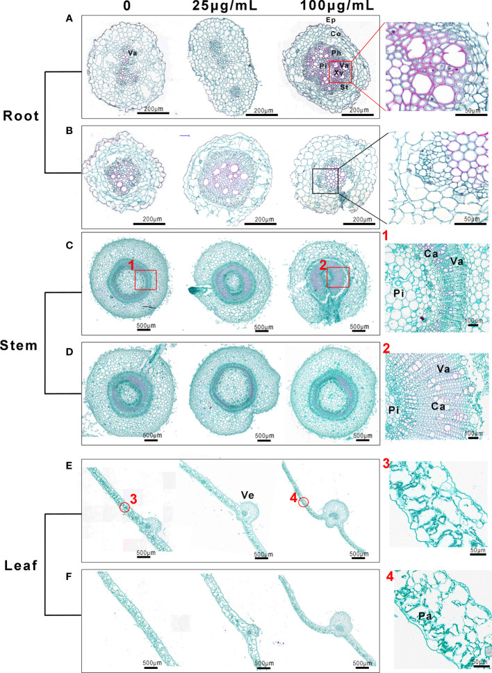
Figure 7.
Morphological structures of tobacco roots, stems and leaves, observed with an optical microscope, after root irrigation (A, C, E) and spraying (B, D, F) with to Mo NPs. The pictures labeled 1 and 2 on the right show the partial magnification of stems in (C) and the pictures labeled 3 and 4 show the partial magnification of roots in (D). Cross-sections of tissues from the same position of seedlings treated for 25 days were used for this assay. Ca, catheter; Co, cortex; Ep, epidermis; Fi, fibers; Pa, palisade tissue; Ph, phloem; Pi, pith; St, sieve tube; Va, vascular bundle; Ve, vein; Xy, xylem;.

