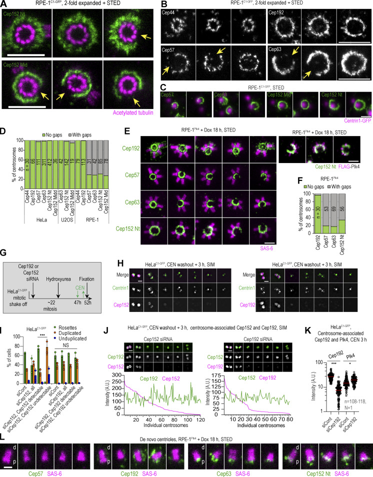
Figure 5.
Asymmetrical distribution of Cep152 in RPE-1 cells and its relationship with Cep192. Logarithmically growing RPE-1C1-GFP cells were immunolabeled for Cep44, Cep192, Cep57, Cep63, or Cep152. Cep152 was detected using an antibody against its N-terminus (Nt) or middle portion (Mid; Fig. S1 A). Some samples were additionally expanded twofold and labeled for acetylated tubulin. (A) The discontinuous appearance of Cep152 signals with gaps (arrows) extending over a variable number of centriole microtubule blades. Centrioles are shown in the top view. (B) The discontinuous appearance of Cep57 and Cep63 showing gaps (arrows). In contrast, Cep44 and Cep192 show continuous ring-like distribution. (C) Cep57, Cep63, and Cep152 signal in relation to Centrin1-GFP signal marking centrioles. Regardless of the gaps in Cep57, Cep63, and Cep152 signals, mother centrioles duplicate and regularly form only one daughter centriole. (D) Quantification of centrosomes with gaps in the signal of the indicated protein around the mother centriole. Centrioles imaged in vertical or near vertical orientation were quantified. (E and F) RPE-1Plk4 cells were treated with Dox for 18 h to induce overexpression of FLAG-Plk4 and formation of centriolar rosettes, and cells were immunolabeled for indicated proteins. (E) Centrosomes with centriolar rosettes to illustrate the absence of procentrioles at regions lacking Cep57, Cep63, and Cep152. Cep192 forms a complete ring around mother centrioles. Right: The absence of Plk4 signal on the regions of the centrosome lacking Cep152. (F) Quantification of centrosomes in RPE-1Plk4 cells exhibiting gaps in the signals of Cep192, Cep57, Cep63, and Cep152. Centrioles imaged in vertical or near vertical orientation were quantified. (G) Experimental strategy used in H–K (for more details, please see Materials and methods). (H) The effect of Cep192 or Cep152 depletion on the assembly of centriolar rosettes. Cells were depleted for Cep192 or Cep152, and centriolar rosettes were induced by transient treatment with CEN and Cep192 and Cep152 were immunolabeled. Centrin1-GFP marks centrioles. (I) Quantification of cells from H containing centriolar rosettes, duplicated centrioles, or at least one unduplicated centriole. 200 cells were counted per condition in each experiment. (J) The effect of Cep192 or Cep152 depletion on Cep152 and Cep192 centrosomal levels. Intensities of Cep192 and Cep152 were determined from widefield recordings and plotted for individual centrosomes. (K) The effect of Cep192 depletion on centrosomal Plk4 levels. Plk4 and Cep192 were labeled and their centrosomal intensity was quantified from widefiled images. Red lines: average intensity. (L) Localization of Cep57, Cep192, Cep63, Cep152, and SAS-6 on de novo–formed centrioles in S phase RPE-1Plk4 cells treated with Dox for 18 h. The wider end of the SAS-6 signal distinguishes the centriole’s proximal (p) end from the distal (d) end. Cep192 associates with the centrioles laterally, Cep63 and Cep152 associate with the centriole’s proximal ends, while Cep57 does not associate with de novo formed centrioles. Scale bars: 0.5 µm for STED, 1 µm for twofold expansion/STED, and 2 µm for widefield.

