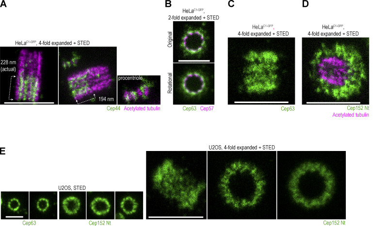
Figure S3.
Spatial arrangement of various centrosomal components. (A–E) Cells were immunolabeled with indicated antibodies and expanded. Some samples were additionally labeled for acetylated tubulin. Centrosomes were imaged using STED. (A) Longitudinally oriented centrosomes immunolabeled for Cep44 and acetylated tubulin. Cep44 localizes to procentrioles from their early stages of formation. (B) STED images of centrioles in top view, co-labeled with Cep63 and Cep57. The two proteins are in proximity, consistent with biochemical analyses showing that they interact. (C) Image of longitudinally imaged centriole immunolabeled for Cep63 signals, showing the distribution of Cep63 in a linear fashion, consistent with its ninefold radial distribution around mother centrioles. (D) Image of a centriole tilted toward the imaging plane and immunolabeled for Cep152 and acetylated tubulin to illustrate the three-dimensional arrangement of the Cep152 signal. (E) Images of Cep63 and Cep152 from U2OS cells show a similar arrangement as found in HeLa cells. Scale bars: 0.5 µm for STED, 1 µm for twofold expansion + STED, and 2 µm for fourfold expansion + STED.

