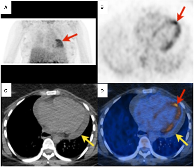
Figure 5.
Myocarditis. Twenty-seven-year-old dyspnoeic woman with widespread concave ST elevation on ECG and increased C-reactive protein (131 mg/L, N < 4), suggestive of myocarditis. 18F-FDG PET revealing diffuse heterogeneous (‘patchy’) myocardial 18F-FDG uptake [red arrows, (A) maximal intensity projection, (B), and (D) axial slices]. Non-enhanced CT showing pericardial effusion [(C), yellow arrow] without 18F-FDG uptake [(D), yellow arrow] related to pericarditis. According to the Bonaca et al.147 criteria, possible myocarditis was retained.

