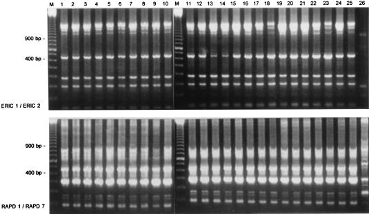
FIG. 2.
RAPD analysis of S. schleiferi strains collected during the present study. The top panel shows results obtained by the combined application of primers ERIC1 and ERIC2; the fingerprints in the bottom panel were generated with the RAPD1-RAPD7 combination. On the left and between lanes 10 and 11 (lanes M), a 100-bp length marker is displayed; fragments with sizes of 900 and 400 bp are highlighted.

