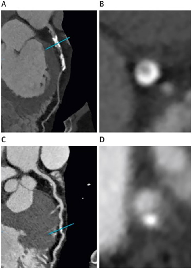
Figure 2:
A) curved multiplanar CT image of atherosclerotic plaque in the left anterior descending artery (LAD). B) cross-sectional image of coronary calcium of the same artery at the level of the blue line in the panel A. C) A curved multiplanar reformatted CT image of LAD of a different patient. D) Cross-sectional image of the same artery at the level of the blue line in the panel C.

