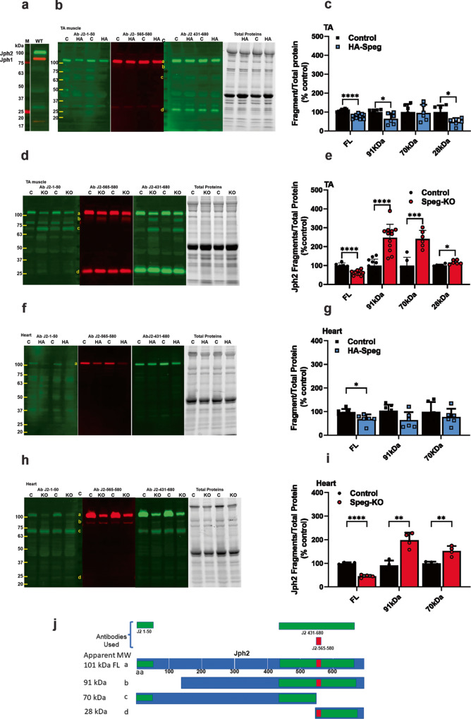
Fig. 4. Effect of Speg deficiency on Jph fragmentation.
a Western blot for Jph2 and Jph1 in the TA of WT mice. Red Jph1. Green Jph2. b Western blot for Jph2 fragments in the TA of control and HA-Speg mice stained with three different antibodies. The antibodies used in the blots are indicated in panel j (see Supplementary Table 3). The far-right panel in (b, d, f, h) is the total protein. c Analyses of fragments in TA of control and HA-Speg mice (n = 6–13). d Western blot for Jph2 fragments in the TA of Speg-KO and control mice. e Analysis of Jph2 fragments in TA control and Speg-KO mice (n = 6–13). f Western blot for Jph2 fragments in hearts of control and HA-Speg mice. g Analyses of fragments in control and HA-Speg hearts (n = 6). h Western blot for Jph2 fragments in control and Speg-KO hearts. i Analysis of Jph2 fragments in hearts of control and Speg-KO mice (n = 4). j Diagram of antibody binding sites and possible calpain-mediated cleavage sites. The tissues used in this experiment were all from male mice are FL- full-length Jph2, b—91 kDa, c—70 kDa, d—28 kDa. Data are plotted as the mean ± SD. ****P < 0.0001, ***P < 0.001, **P < 0.01, and *P < 0.05.

