. 2023 Jun 30;29(18):3813–3825. doi: 10.1158/1078-0432.CCR-23-0827

Figure 6.

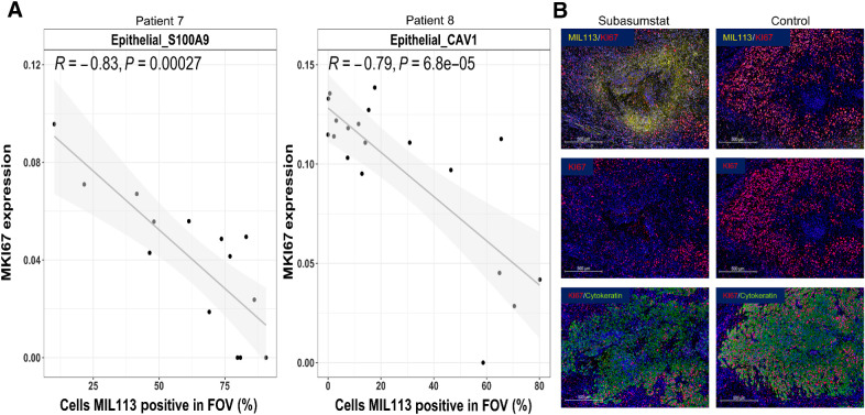
Figure 6. Subasumstat target engagement is specifically associated with a reduction in markers of cell-cycle progression in malignant epithelial cells from patients 7 and 8. A, Correlation of MKI67 expression to drug target engagement measured by the percentage of cells MIL113-positive in Epithelial_S100A9 cells (patient 7) and Epithelial_CAV1 cells (patient 8), respectively. Linear models were fit and are shown with associated Pearson correlation, P value, as well as 95% confidence intervals (shaded). B, Immunofluorescent staining for subasumstat-SUMO adducts (MIL113, yellow), cell proliferation marker KI67 (red), and tumor cell marker (cytokeratin, green) showing the reduction in proliferating tumor epithelium in regions of subasumstat drug target engagement compared with a directly adjacent uninjected region, no drug control; scale bar, 500 μm.

Subasumstat target engagement is specifically associated with a reduction in markers of cell-cycle progression in malignant epithelial cells from patients 7 and 8. A, Correlation of MKI67 expression to drug target engagement measured by the percentage of cells MIL113-positive in Epithelial_S100A9 cells (patient 7) and Epithelial_CAV1 cells (patient 8), respectively. Linear models were fit and are shown with associated Pearson correlation, P value, as well as 95% confidence intervals (shaded). B, Immunofluorescent staining for subasumstat-SUMO adducts (MIL113, yellow), cell proliferation marker KI67 (red), and tumor cell marker (cytokeratin, green) showing the reduction in proliferating tumor epithelium in regions of subasumstat drug target engagement compared with a directly adjacent uninjected region, no drug control; scale bar, 500 μm.