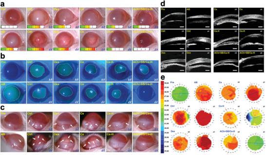
Figure 5.

Clinical observations. a) Slit‐lamp biomicroscopic, b) corneal fluorescein staining, c) lateral view slit‐lamp biomicroscopic, d) ultrasound biomicroscopic, and e) corneal topography images of alkali burn (AB) eyes at 8 h and 4 d post‐instillation of ATS (Ctrl), dexamethasone eye drop (Dex), and ACh+SB/Ce‐H nanoformulation and its components including SRCNs (Ce) and poly(l‐histidine) functionalized SRCNs (Ce─H), respectively. Scale bars in d) are 500 µm. The score gauge in a) is the grade of corneal haze. A dotted white line in b) marks the margin of wound; white arrow in c) represents abnormal blood vessels.
