Figure 6.

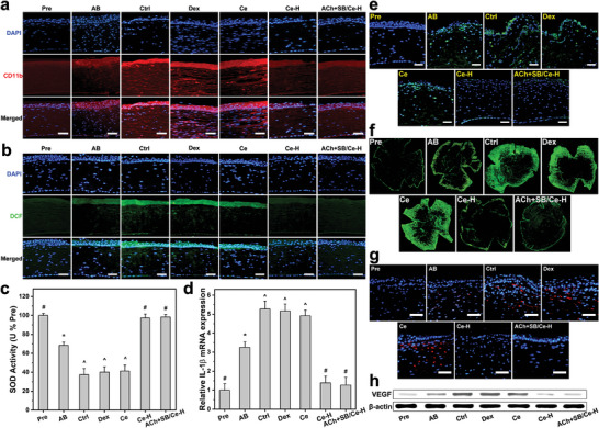
Therapeutic activity assays at 4 days post‐instillation. Immunofluorescence images of a) CD11b (red fluorescence)‐ and b) DCF (green fluorescence)‐stained corneas of AB rat eyes instilled with ATS (Ctrl), dexamethasone eye drop (Dex), Ce, Ce‐H, and ACh+SB/Ce‐H, respectively. c) SOD activity and d) IL‐1β levels in the corneal tissues of the treated AB eyes. e) Fluorescence images of different corneal sections stained with TUNEL (green fluorescence). f) Fluorescence images of flat‐mount whole corneal tissues; blood vessels are visualized by CD31 staining (green fluorescence). g) CD31 (red fluorescence) immunofluorescence staining images of different test corneal tissues. h) Western blot analysis of VEGF expression. Scale bars: 50 µm. Values are mean ± SD (n = 10); *P < 0.05 versus all groups; # P < 0.05 versus AB, Ctrl, Dex, and Ce groups; ^ P < 0.05 versus Pre, AB, Ce─H, and ACh+SB/Ce─H groups. The inclusion of the healthy (Pre) and as‐induced AB (AB) groups in all panels aims for comparative studies.
