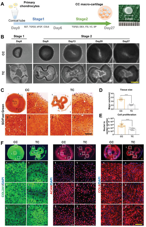
Figure 3.

CC‐chons can form large‐size and ECM‐rich macro‐cartilage in 3D culture. A) Schematic diagram illustrating the designed program for step‐wise pellet culture. B) Representative bright‐field images of tissues by CC and TC incubation over time. Scale bar: 1 mm. C) SO/fast green staining of CC and TC tissues. Scale bar: 100 µm. D) Major axis quantification of center section in CC and TC tissues (n = 3, unpaired two‐tailed Student's t‐tests). E) Quantification of cell number in the central section (n = 3, unpaired two‐tailed Student's t‐tests). F) Representative images of immunofluorescent staining of chondrocyte markers (COL2A1, ACAN, and COL6) in CC and TC tissues. Scale bar: 100 µm. All data were mean ± SEM. ****p < 0.0001.
