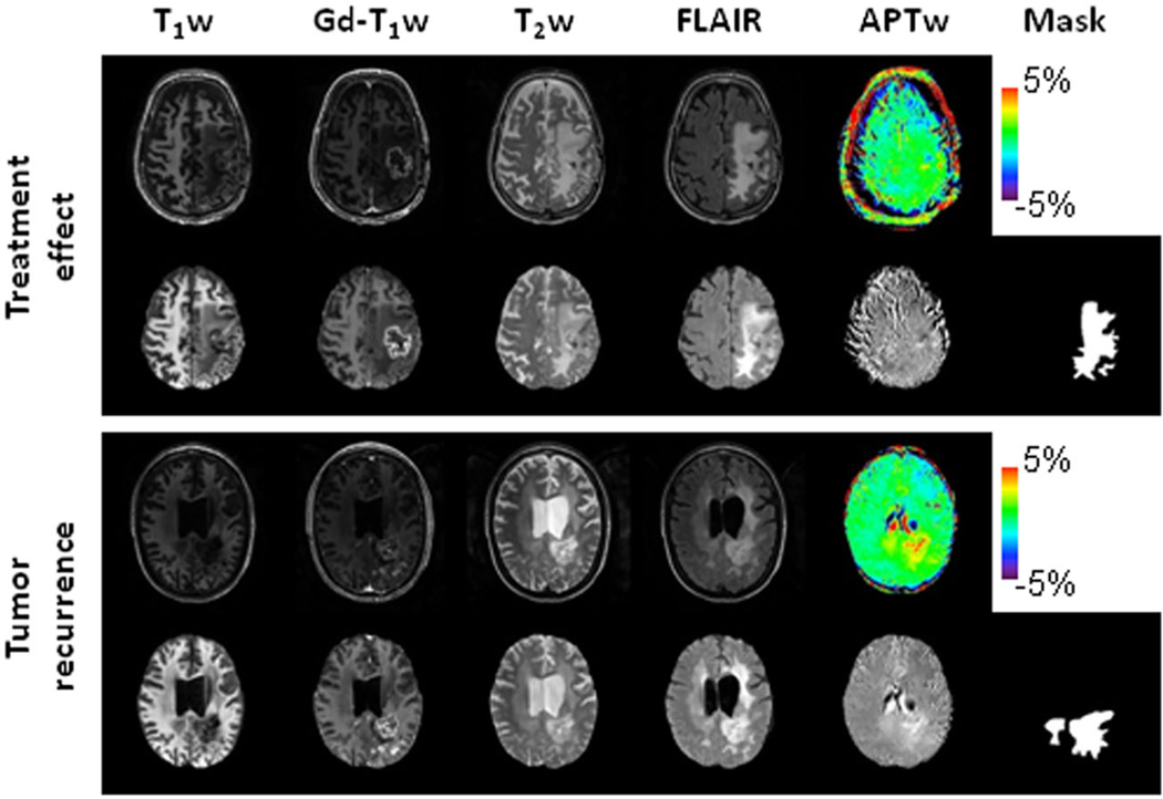
FIGURE 2.
Structural and APTw MR images before and after image preprocessing, as well as masks for two post-treatment patients. Top case, a glioblastoma patient (male, 65 years) with treatment effect. The lesion showed a homogeneous isointensity to minimal hyperintensity on the APTw image. Bottom case, an anaplastic astrocytoma patient (female 42 years) with tumor recurrence. The lesion showed a strong hyperintensity on the APTw image. In this case, the cerebrospinal fluid in ventricles presented some artifact (APTw hyperintensity). Notably, the masks covered only the tumor and tumor-associated vasogenic edema. To focus on the direct effects related to the tumor, the masks did not include the periventricular edema in the bilateral frontal lobes, which were detached from the original location (bottom case).

