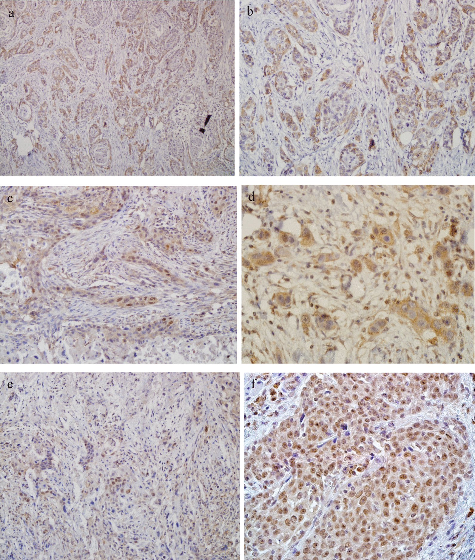
Fig. 1.

Immunohistochemistry staining of B19V VP1/VP2 antigens, NF-κB p65, and p16INK4a in moderately differentiated HNSCC with hematoxylin counterstain. a, b Represent positive B19V VP1/VP2 staining in the cytoplasm of many malignant HNSCC cells with 100× and 200× magnifications, respectively. c, d Represent nuclear and cytoplasmic NF-κB p65 immunostaining in HNSCC cells with the magnifications of 200× and 400×, respectively. e, f Indicate nuclear and cytoplasmic p16INK4a immunostaining in HNSCC cells with 200× and 400× magnifications
