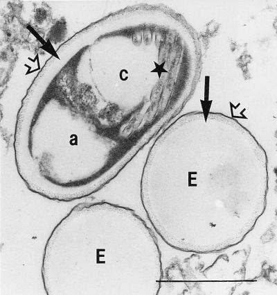
FIG. 8.
Microsporidian spores from human stool samples as shown by light microscopy in Fig. 3 and 4. Two spores are empty looking (E). One spore shows a distinct compartmentation built up of two electron-lucent areas (a and c, with cross sections of the polar tube [★] shown in area c) separated by an electron-dense area. All three spores reveal the bilayered spore wall consisting of an outer electron-dense layer (exospore) (➩) and an inner electron-lucent layer (endospore) (➞). Bar, 1 μm. Magnification, ×30,000.

