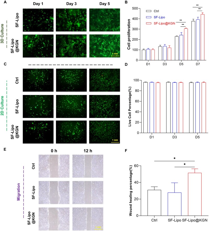
Figure 2.
SF-Lipo@KGN scaffolds exhibited excellent biocompatibility. (A) Fluorescence image of DIO-labeled chondrocytes growing on a 3D scaffold. Scale bar = 1 mm. (B) CCK-8 was used to measure the effect of scaffolds on cell proliferation. (C, D) The survival of cells co-cultured with scaffold was determined using the live/dead staining. Scale bar = 1 mm. (E, F) Wound healing experiment to verify the effect of KGN on chondrocyte migration. Scale bar = 1 mm. The data are presented as mean ± SD (n = 3). *Indicates statistically significant differences where P < 0.05, **where P < 0.01.

