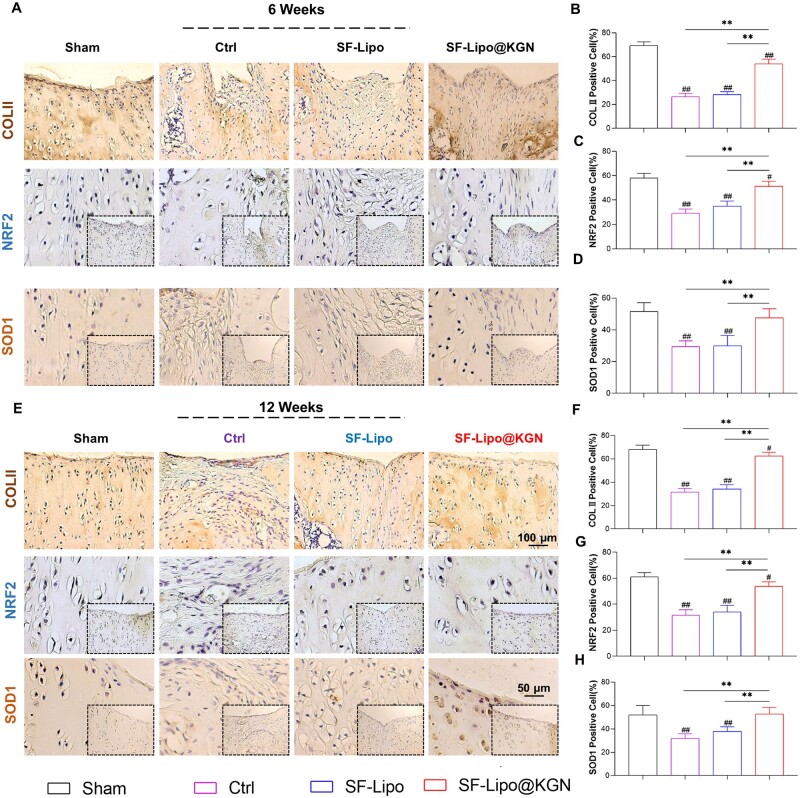
Figure 6.
The SF-Lipo@KGN scaffold promotes the expression of matrix protein and antioxidant protein in cartilage tissue. (A) Representative IHC images of cartilage defects stained with COLII, NRF2 and SOD1 at 6 weeks after surgery. Scale bar = 100 μm and 50 μm. (B–D) Quantifying the percentage of COLII-positive, NRF2-positive, and SOD2-positive cells in the articular cartilage at 6 weeks after surgery. (E) Representative IHC images of cartilage defects stained with COLII, NRF2 and SOD1 at 12 weeks after surgery. Scale bar = 100 μm and 50 μm. (F–H) Quantifying the percentage of COLII-positive, NRF2-positive, and SOD1-positive cells in the articular cartilage at 12 weeks after surgery. The data are presented as mean ± SD (n = 6). *Indicates statistically significant differences where P < 0.05, ** where P < 0.01 between the indicated groups; # where P < 0.05, ## where P < 0.01 versus the sham group.

