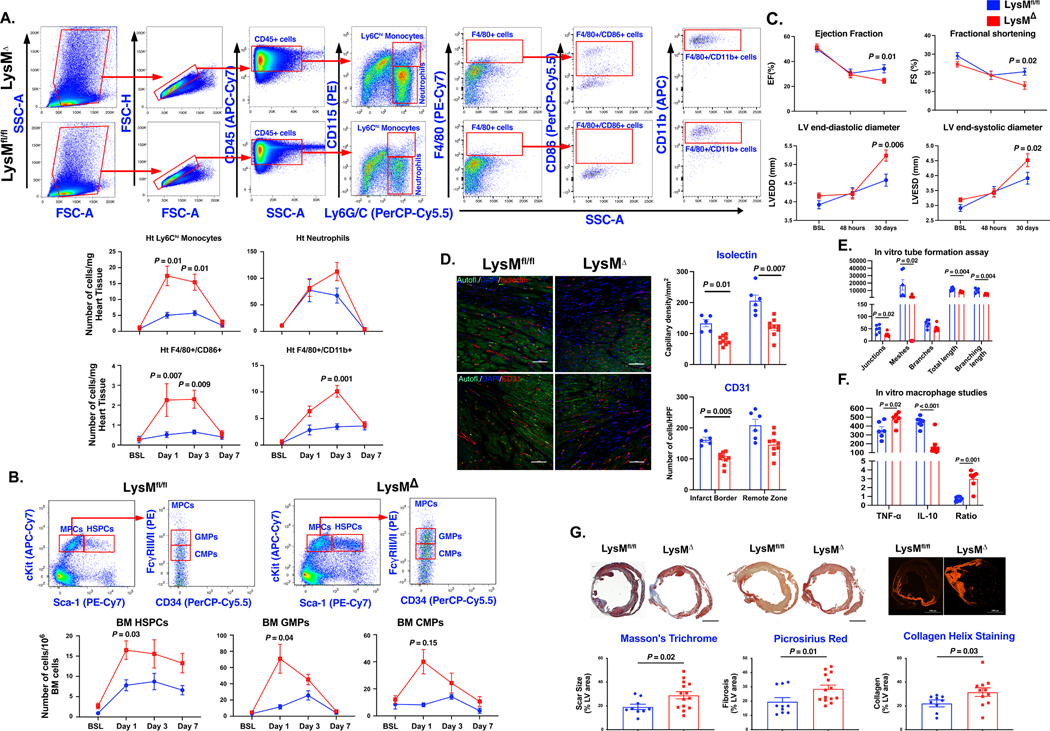
Figure 1. Myeloid deficiency of Plpp3 aggravates cardiac inflammatory response following acute myocardial ischemia.
A, Representative flow cytometry plots and analyses of cardiac inflammatory cells demonstrating elevated Ly6Chi monocytes, pro-inflammatory macrophages, and neutrophils, in LysM-Plpp3Δ mice after myocardial infarction (n=6–8 mice/group/time-point). B, Significant increase in the number of bone marrow HSPCs (Sca-1+/c-Kit+/Lin−), CMPs (Lin−/c-Kit+/Sca-1−/CD16/32−/CD34+), and GMPs (Lin−/c-Kit+/Sca-1−/CD16/32+/CD34+) in LysMΔ mice (n=6–8 mice/group/time-point). C, Echocardiography demonstrates significant deterioration in LV function and remodeling in LysM-Plpp3Δ (n=10–15 mice/group). D, Lower capillary density in mice with myeloid-specific LPP3 deletion (n=5–9 mice/group, scale bars represent 50 μm). E, HUVEC cells showing significant reduction in multiple parameters of tube formation assay when treated with LysM-Plpp3Δ BMDM supernatant (N = 3–5 technical repeats). F, LPS-stimulated LysM-Plpp3Δ BMDM exhibit exacerbated inflammatory response as demonstrated by the higher levels of TNF-α and lower expression of IL-10 (N = 3–5 technical repeats). G, Representative images of Masson’s trichrome, Picrosirius red staining, and Collagen Hybridizing Peptide Cy3 Conjugate (R-CHP) staining (upper panel) performed 30 days after LAD ligation, demonstrating a significant increase in scar size and fibrosis in LysM-Plpp3Δ mice (n=9–14 mice/group/analysis, scale bar = 2 mm). Throughout the figure, data are represented as mean ± SEM. Repeated analyses were conducted using repeated measures ANOVA, the Geisser-Greenhouse correction for unequal variance, and the Sidak posthoc test. Comparison between 2 groups was performed using the Mann-Whitney test.

