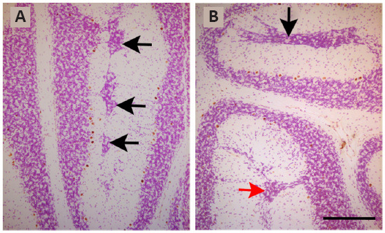
Figure 2.

Light photomicrographs illustrating ectopic granule cells from rats administered with bromodeoxyuridine on embryonic days 13–14 and sacrificed on postnatal day 90.
Black arrows in “A” display ectopic neurons at the bottom of the prima fissure. Black and red arrows in “B” indicate ectopic granule cells at the bottom of the prima fissure and secunda fissures, respectively. Scale bar: 300 μm. Unpublished data.
