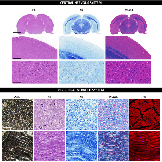
Figure 2.

Histochemical staining techniques in central and peripheral nervous systems.
Representative coronal sections of rat encephalus, and cross and longitudinal sections of rat sciatic nerves stained by diverse histochemical methods. Here a comparison between the routine stain HE and specific myelin staining methods such as KB (or conventional LFB), MCOLL, OsO4 and FM staining is included. HE shows the typical histological pattern with nuclei stained blue and a pink contrast. Conventional KB reveals the myelin sheaths with an intense blue histochemical reaction and cellular elements in a light purple color. The MCOLL method provides a simultaneously and specific staining of myelin (blue) and fibrillar collagen fibers (red) with a cell nuclei contrast (blue/purple). OsO4 generates a permanent and stable myelin dark stain, which significantly facilitates the identification and evaluation of nerve fibers. FluoroMyelin™ stains myelin in red fluorescent color and nuclei were detected by DAPI (blue). All samples were fixed in formaldehyde and embedded in paraffin except FluoroMyelin™ ones, which were fixed in formaldehyde, cryopreserved and cryosectioned. Scale bars in images for central nervous system: low magnification: 3000 μm; medium magnification: 500 μm; higher magnification: 50 μm. Scale bars in images for peripheral nervous system: 50 μm cross and longitudinal sections. DAPI: 4′,6-Diamidino-2-phenylindole; FM: FluoroMyelin™; HE: hematoxylin and eosin; KB: Klüver-Barrera; LFB: Luxol fast blue; MCOLL: myelin-collagen method; OsO4: osmium tetroxide.
