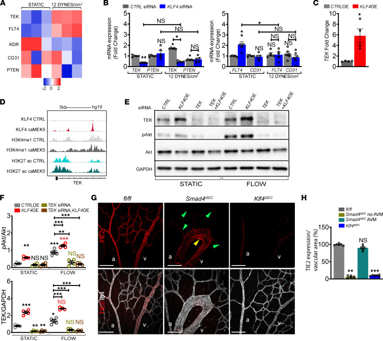
Figure 6. KLF4-mediated TEK expression is required for flow-induced PI3K/Akt activation.
(A) Heatmap of potential mediators of PI3K signaling in CTRL siRNA HUVECs grown in static versus subject to 12 DYNES/cm2; n = 3/group. Color key shows log2 change upon FSS stimulation. (B) qPCR for TEK and PTEN (left panel) and FLT4 and CD31 (right panel) in CTRL versus KLF4 siRNAs HUVECs grown in static or subject to 12 DYNES/cm2 (n = 5/group). (C) TEK mRNA expression in CTRL-OE versus KLF4-OE HUVECs (n = 4/group). (D) Reanalysis of previous published CHIP-Seq data of KLF4 overexpression (caMEK5) in primary human pulmonary artery endothelial cells (PAEC) with the Integrative Genomics Viewer (IGV). Two distinct peaks within enhancer regions of the TEK gene were identified. (E) WB for indicated proteins of HUVECs transfected with CTRL and TEK siRNAs and CTRL-OE and KLF4-OE constructs grown in static or subject to 12 DYNES/cm2 for 4 hours. (F) Quantification of pAkt/Akt and TEK/GAPDH in indicated genotypes (n = 4/group). (G) Labeling of Tx-induced P6 fl/fl, Smad4iΔEC and Klf4iΔEC retinas with anti-TIE2 antibody (red) and IB4 (white). Green and yellow arrowheads indicate non-AVM versus AVM region, respectively. (H) Quantification of TIE2 expression per vascular area in the indicated genotypes (n = 6 [2 images/retina]/group). a, artery; v, vein. Scale Bars: 100μm in G. Mann-Whitney test (C) and 1-way Anova (B, F, and H) were used to determine statistical significance.Data are represented as mean ± SEM. *P < 0.05, **P < 0.01, ***P < 0.001.

