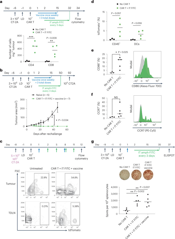
Fig. 5. Amph-FITC therapy induces epitope spreading to elicit an endogenous antitumour T cell response.
a, Flow cytometry on treated CT-2A tumours on day 34 after adoptive transfer of CD45.2+ FITC CAR T cells, quantifying CD4+ versus CD8+ CD45.1+ tumour-infiltrating host T cells (n = 4 animals per group). b, CT-2A tumour-bearing mice previously cured with CAR T and amph-FITC therapy were rechallenged versus naïve mice with 106 CT-2A cancer cells on the opposite flank at day 92 following adoptive transfer (n = 5 animals per group). c,d, Representative flow plots of DCs (c) in tumours and TDLN of mice bearing tdTomato+ CT-2A tumours at 12 days post-adoptive transfer and quantification (d) of tdTomato+ DCs in TDLN (n = 3 animals per group for no CAR T group, n = 4 for CAR T + i.t. FITC group). e,f, Expression of activation markers CD86 (e) and CCR7 (f) in TDLN DCs at 12 days post-adoptive transfer with representative histograms (n = 3 animals per group for no CAR T group, n = 5 for CAR T + i.t. FITC group). g, ELISPOT on spleens of CT-2A tumour-bearing mice on day 37 post adoptive transfer (n = 5 animals per group). Splenocytes were stimulated with irradiated CT-2A cancer cells. All replicates are biological replicates. P values were determined by unpaired Student’s t-test. Error bars represent s.d. NS, not significant; *P < 0.05, **P < 0.01.

