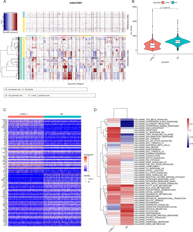
Fig. 6.
Epithelial cells in brain metastases are more malignant. A Copy number variation in Epithelial cells, endothelial cells, and fibroblasts were used as a reference, red represents overexpression of genes, and blue represents low expression. B Violin plot indicating the CNV scores of Epithelial cells from lung adenocarcinoma and brain metastases. C Heatmap showing the top 50 differential expressed genes between the two groups. D Differences in pathway activities by GSVA between lung adenocarcinoma and brain metastases epithelial cells

