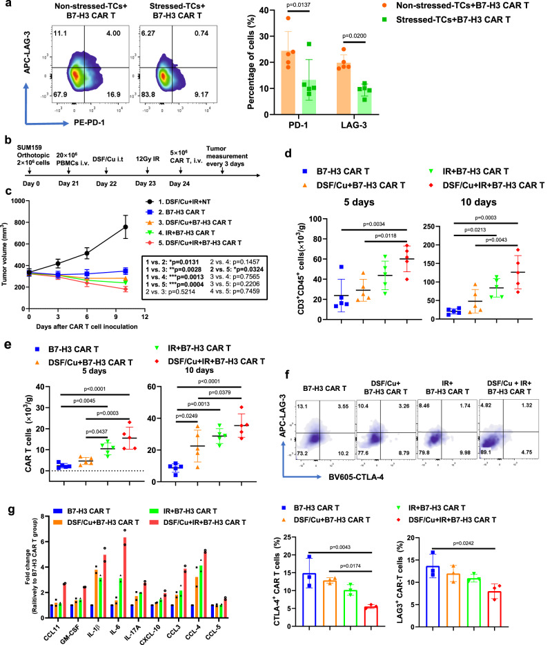
Fig. 5. Tumors stressed by DSF/Cu and IR reverse immunosuppressive TME in humanized mice.
a Exhausted B7-H3 CAR T cells (CD3+PD-1+ or CD3+LAG-3+) after round 3 (R3) of repetitive co-culture with DSF/Cu+IR-stressed vs. non-stressed PANC-1 cancer cells (n = 5 independent experiments). b Schema of the humanized mouse tumor model. c Tumor volumes (n = 5 mice/group) in the humanized mice were measured every 3 days. d Number of the total human CD3+ CD45 + cells including engrafted human PBMC and CAR T cells in tumor tissues 5 days and 10 days after CAR T cell infusion (n = 5 mice/group). e Number of B7-H3 CAR T cells in tumor tissues 5 days and 10 days after CAR T cell injection (n = 5 mice/group). f B7-H3 CAR T cells expressing markers associated with T-cell exhaustion (CD3+CD45+LAG-3+ or CD3+CD45+CTLA-4+) in tumor-infiltrating CAR T cells in tumor tissues 10 days after CAR T cell infusion (n = 5 mice/group). g Cytokine and chemokine levels in tumor tissues on day 5 after CAR T cell injection (Tumor homogenate was pooled to yield 2 samples from 5 mice/group). Statistical comparisons were performed using two-tailed unpaired t-test (a), two-way ANOVA with Tukey’s multiple comparisons test (c), and one-way ANOVA with Tukey’s multiple comparisons test (d, e, f). P-values are shown and error bars indicate mean ± SD. Source data are provided as a Source Data file.

