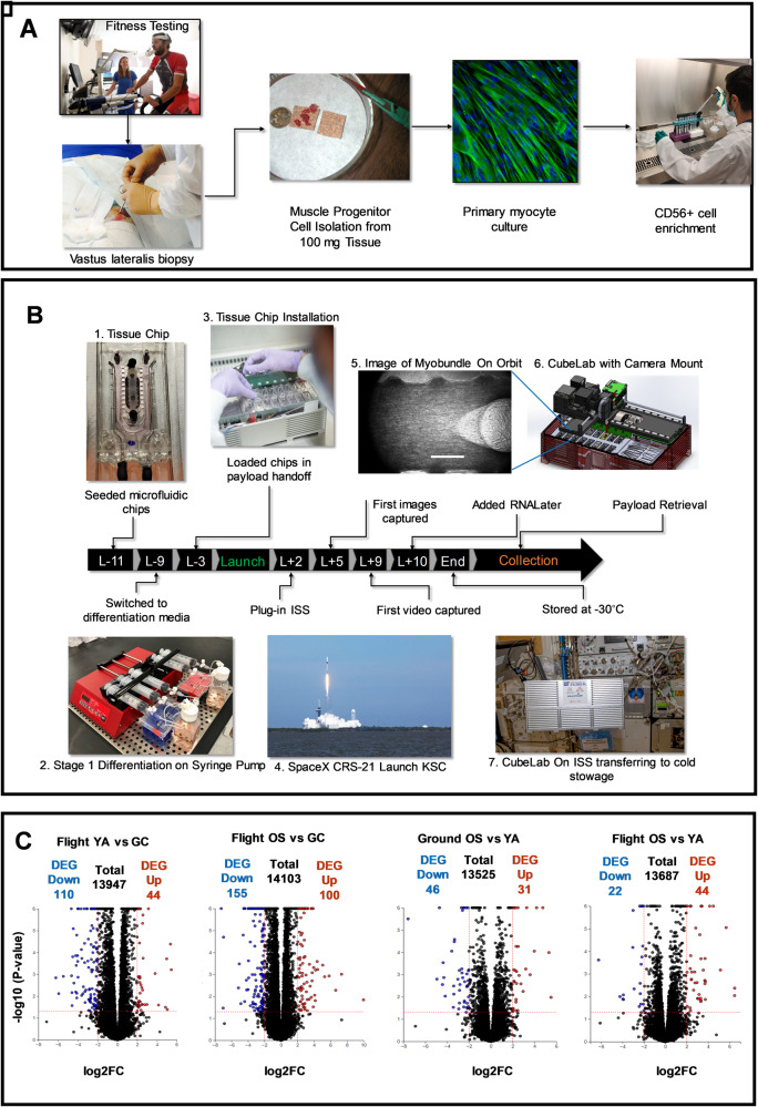
Fig. 1. Experimental design, implementation, and RNA analysis of the skeletal muscle MPS payload for the international space station.
A Human cell banks. Skeletal muscle biopsies were obtained from the vastus lateralis from volunteers (AdventHealth, Orlando). Isolated muscle precursor cultures were enriched for CD56+ (myogenic) cells. B Experimental flight timeline. Photos are numbered in the order of the process. 1. Tissue chips were seeded with myoblasts, 2. pre-differentiated using a syringe pump, 3. loaded into the CubeLab™ and 4. launched to the ISS on SpaceX CRS-21. Crew members installed the PAUL on the EXPRESS rack locker and the on-orbit experiment was initiated after plug-in. 5. Images of the myobundles were captured on orbit by the optics system installed into the lid of the CubeLabTM. Scalebar = 500 μm. 6. The Optics system automatically moved in the x,y,z axes to collect images. 7. Ten days post launch, crew members moved the payload to cold stowage following experiment termination with RNALater. C RNA-Seq volcano plots. YA flight vs ground; OS flight vs ground; Ground OS vs YA and Flight OS vs YA. Colored points are differentially expressed between comparisons. Blue: significantly down-regulated genes. Red: significantly up-regulated genes. Significance is determined according to log2 fold change with threshold set at ±2 and -log10 p-value ≤ 0.05. Vertical dotted lines are positioned at log2 fold-change of ±2 and horizontal dotted lines are positioned at the -log10 p-value = 0.05. RNA-Seq analysis measured three distinct chips each containing one myobundle derived from five male donor cells pooled in equal ratios.

