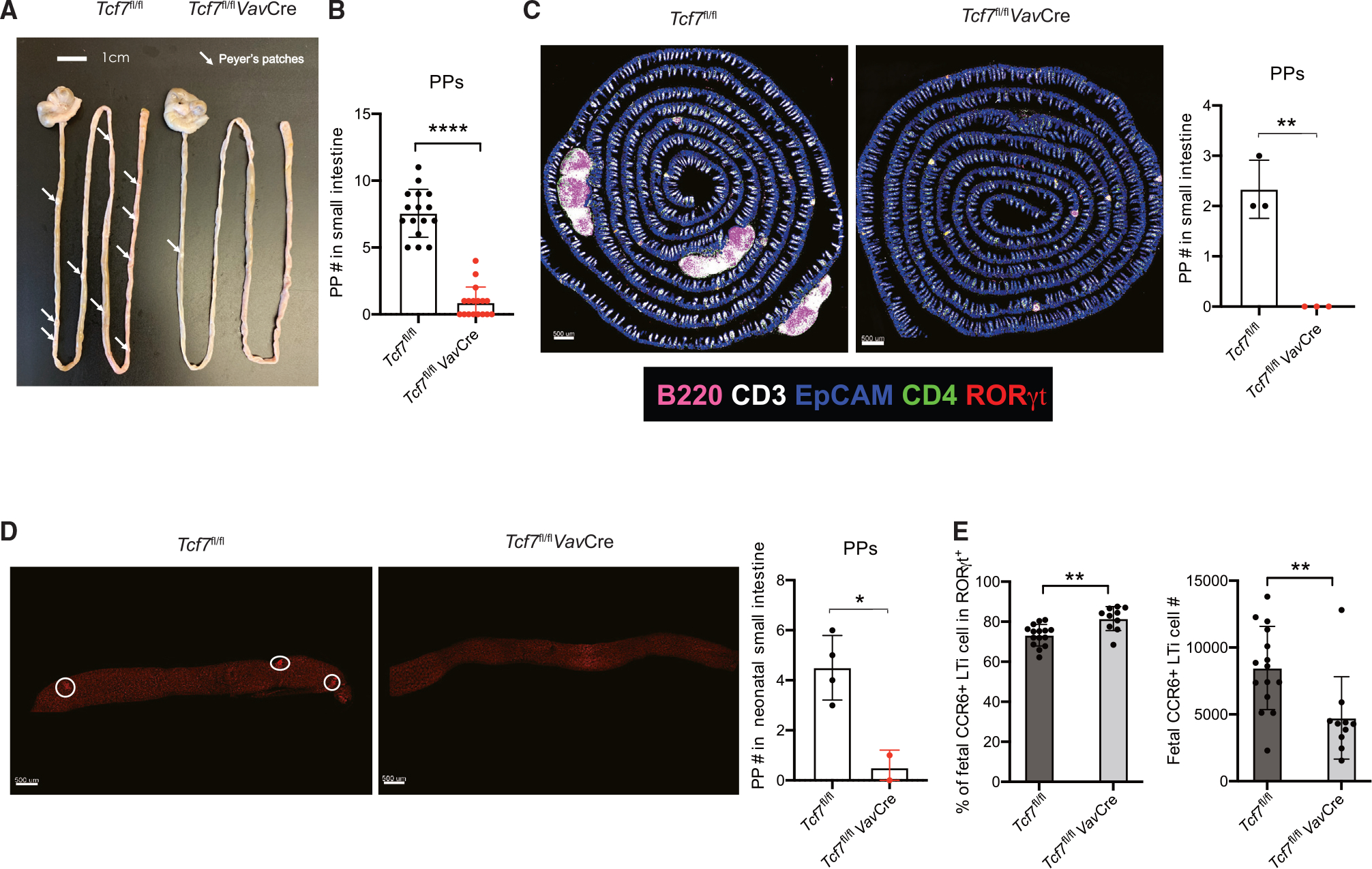
Figure 3. TCF-1 is required for the formation of PPs, but not ILFs, in the small intestine.

(A) Visualization of PPs from WT and Tcf7fl/flVavCre mice.
(B) Quantification of PPs from the mice in (A). Each symbol represents an individual mouse.
(C) Immunofluorescent staining of PPs in small intestine (left) from 6- to 8-week-old mice. Quantification of PPs (right). Each symbol represents an individual mouse.
(D) Visualization of PPs from neonatal (day 1) mice by immunofluorescent staining of RORγt. Each symbol represents an individual mouse.
(E) Cells were isolated from Tcf7fl/fl and Tcf7fl/flVavCre fetal (E16.5) intestine, and CCR6+ LTi population was analyzed; bar graphs show quantification in percentages and total cell numbers. Each symbol represents an individual mouse or fetus. mean ± SD; n = 15–16 in (B), 2–4 in (C)–(D), and 10–15 in (E); *p < 0.05, **p < 0.01, ****p < 0.0001, Student’s t test. Data are representative of at least three independent experiments. See also Figure S3.
