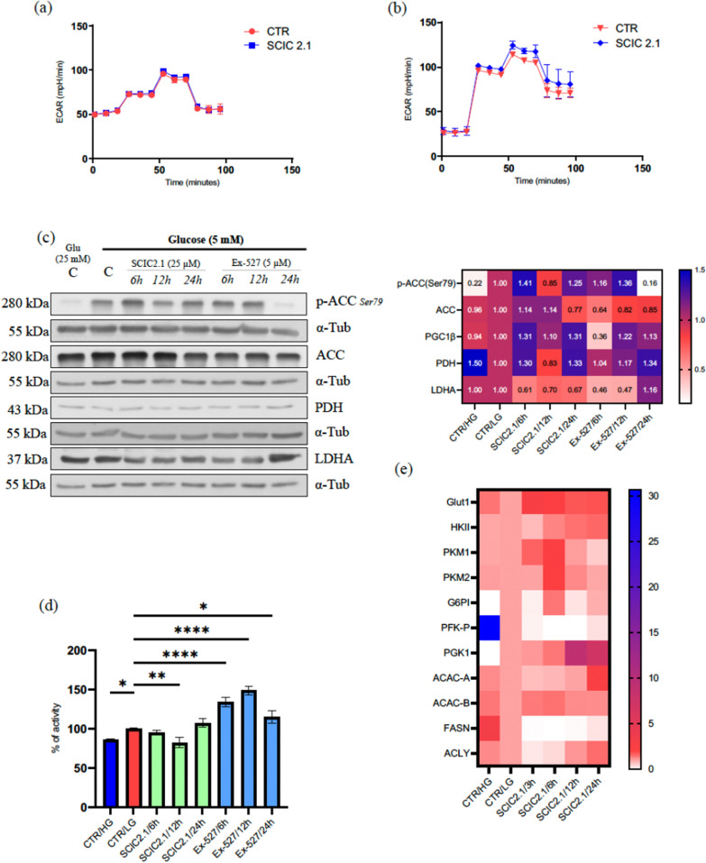
Fig. 5.
SCIC2.1 alters the metabolic program and inhibits lipogenesis upon glucose starvation. Extracellular acidification Rate (ECAR) of HepG2. a Glucose (25 mM) exposure followed by the treatment of SCIC2.1 b Glucose (5 mM) exposure followed by the treatment of SCIC2.1. c Western blot analyses of SCIC2.1 alters the metabolic program upon glucose starvation: Western blot analyses p-ACC (Ser79), ACC, PDH, and LDHA was performed in HepG2 cells treated with SCIC2.1 and Ex-527 at indicated concentration and time points. Heat map showing the band quantification of protein expression. d quantification of Oil Red Oil staining at 540 nm (n = 3) using TECAN in HepG2 cells treated with SCIC2.1 and Ex-527 at indicated time points. e Heat map showing different expression levels of genes involved in glycolysis and lipid metabolism in HepG2 cells treated with SCIC2.1 at 25 µM. Statistical significance was calculated using the student t-test or one-way ANOVA using GraphPad Prism 9.4.0, and statistical significance is expressed as *p-value < 0.05, **p-value < 0.01, and ****pvalue < 0.0001 vs control. Error bars represent the standard deviation (SD) of three biological replicates

