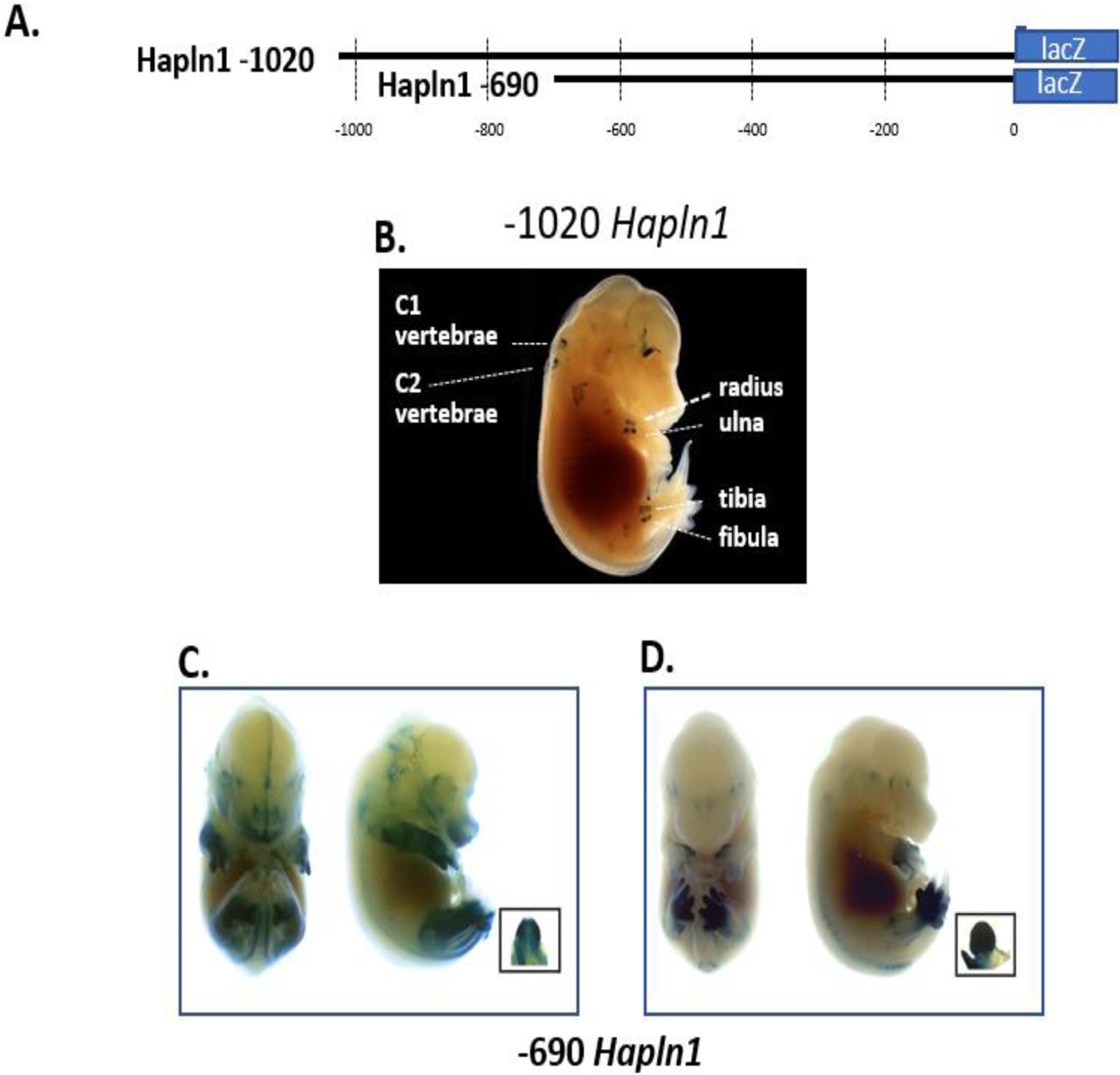
Fig 1.

Link protein gene promoter constructs show in vivo expression patterns in either the skeleton or limb/genitalia. (A) Two different link protein promoter fragments consisting of the −1020 and −690 nt regions were tested in transgenic mice. (B) A representative E15.5 mouse transiently expressing the −1020 promoter region showed only X-gal staining in appendicular and axial cartilaginous skeletal tissues. A dark background was used to provide contrast to the photograph to allow easy visualization of the discrete expression in cartilage sites. (C) and (D) The shorter −690 promoter region demonstrated high-level expression in the limb and genitalia as shown in two representative mouse embryos. As shown, embryos showed variability of X-gal staining in the limb and hind paw regions.
