
Figure 1. Acquisition and segmentation of brain structural images in infants with CAH and age-matched controls for volumetric analyses.
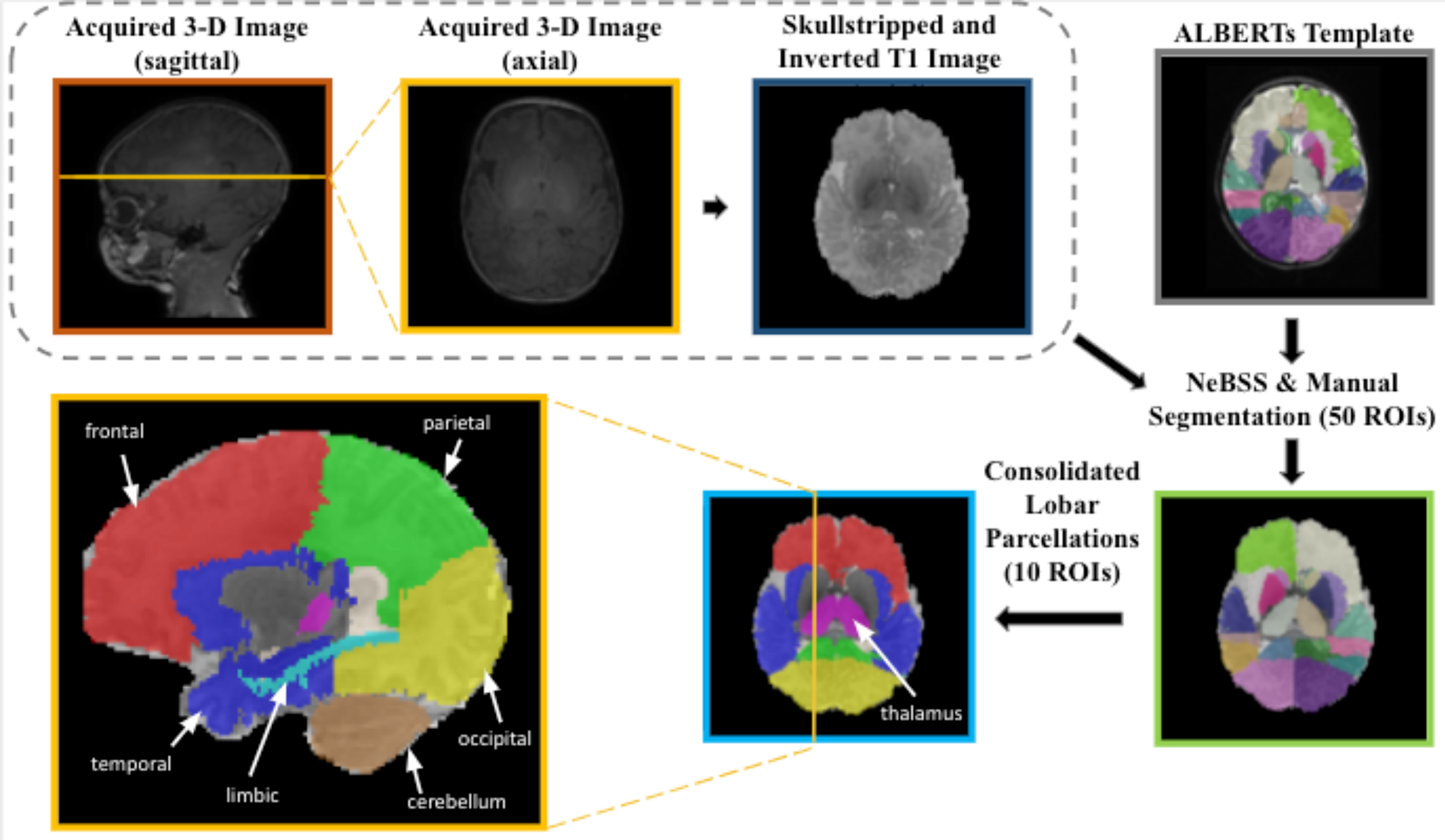
Prior to automated segmentation, 3D T1-weighted anatomical images were automatically skull-stripped (FSL) and then edited manually (ITK-SNAP) and inverted into pseudo-T2-weighted images to match the imaging modality of the ALBERTs atlases. The images were then bias-corrected and automatically segmented (NeBSS) into 50 brain tissue types. These 50 subregions were then refined into larger structural regions of interest (ROIs).