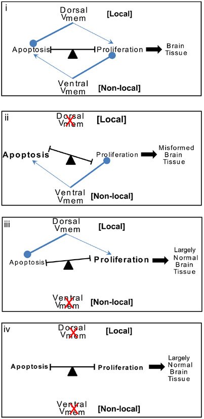
Fig. 6. A model for Vmem regulation of apoptosis-proliferation balance towards shaping brain morphology.
Model for Vmem regulation of brain tissue sculpting. Both dorsal and ventral Vmem signals are involved in brain tissue sculpting by their effects on the balance of apoptosis and proliferation within the tissue. Under normal conditions (i) the dorsal Vmem strongly inhibits apoptosis and mildly stimulates proliferation locally within the developing brain. The ventral Vmem strongly inhibits proliferation and mildly stimulates apoptosis at a distance in the developing brain tissue. Coordination of these two Vmem signals results in proper balance of proliferation-apoptosis and ultimately sculpting of the brain tissue during development. Blocking of dorsal Vmem signal (ii) releases the strong inhibition on apoptosis and further facilitates strong inhibition of proliferation by the ventral Vmem signals. This results in a shift in balance towards apoptosis resulting in malformed brain tissue. Blocking of ventral Vmem signal (iii) releases the strong inhibition on proliferation and increases the inhibition of apoptosis by the dorsal Vmem signal. This results in significant increase in proliferating cells but otherwise largely normal brain structure development. Blocking of both dorsal and ventral Vmem signal (iv) cancel each other’s effect on proliferation and apoptosis leaving the sculpting of brain to other regulators of brain structure development resulting in largely normal brain structure.

