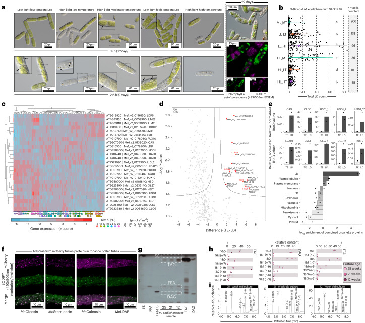
Fig. 6. LDs accumulate in Mesotaenium upon changing environments.
a, DIC and confocal micrographs of Mesotaenium endlicherianum SAG 12.97 cells accumulating LDs (arrows) upon exposure to different temperature/light conditions (abbreviations) of the gradient table for 89 h or 216 h. For confocal microscopy, algae were cultured independent of table conditions at 75 µmol photons m−2 s−1 and 22 °C for 22 days. LDs are visible as distinct globular structures and were stained with BODIPY (false-coloured green; 493 nm excitation, 503 nm emission); chlorophyll autofluorescence in false-coloured purple; for each condition, at least ten micrographs were taken, all showing similar phenotypes of the cells. b, Violin plots of LD quantification after 9 days of exposure to different environmental conditions; significance grouping (Mann–Whitney U) is based on P < 0.05; see also Supplementary Fig. 27. c, Heat map of row-scaled z scores of the expression of homologues for LD biogenesis and function (see also Supplementary Fig. 28). Conditions are displayed at the bottom as symbols in different colours; best Arabidopsis hits (via BLASTp) are shown on the right. d,e, Proteomic investigation into lipid-enriched phases extracted from Mesotaenium; note the enrichment in hallmark proteins of LDs. Volcano plot showing significantly (false discovery rate (FDR) <0.05) enriched Mesotaenium proteins in the lipid-enriched (LD) versus the TE (d). Bar plots show the relative, normalized iBAQ values for ten LD signature proteins detected in Mesotaenium (e). Bottom bar plot shows the log2 enrichment of proteins characteristic for subcellular compartments. LL, ML and HL, low, moderate and high light; LT, MT and HT, low, moderate and high temperature, respectively; ER, endoplasmic reticulum. f, LD proteins of M. endlicherianum localize to LDs in tobacco pollen tubes: cLSM images of transiently expressed proteins appended to mCherry in transiently transformed N. tabacum pollen tubes. LDs were stained with BODIPY 493/503; for each construct, the images are representative of at least nine micrographs of transformed pollen tubes per fusion construct. Scale bars, 10 µm. g,h, Lipid composition in M. endlicherianum LDs of 12- to 25-week-old cultures and standards for sterol esters (SE), FFA, free sterols (free S), TAG and DAG via analytical TLC (g) and preparative TLC followed by GC for profiling (h). i, Full lipid profiles assessed via GC.

