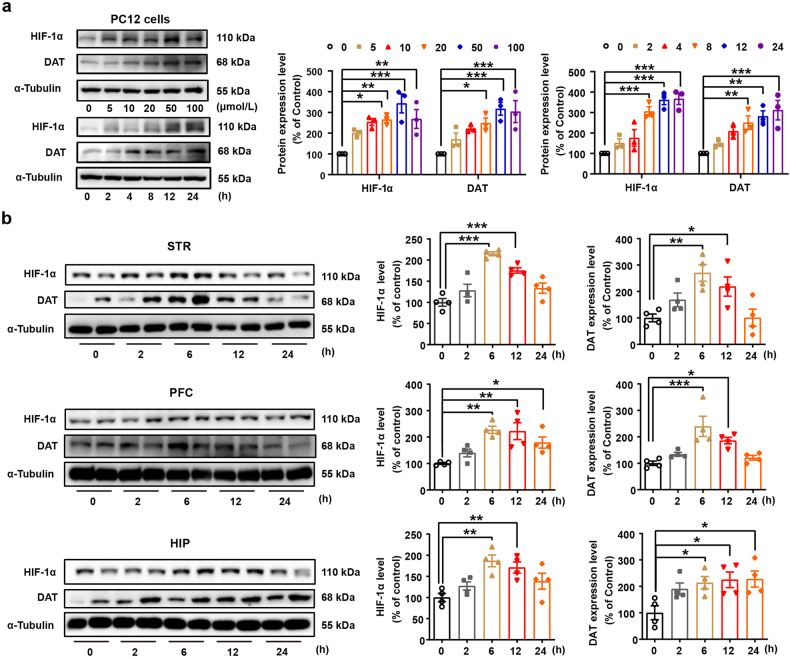
Fig. 4.
Rox treatment increases dopamine transporter (DAT) expression. a PC12 cells were treated with roxadustat 24 h at indicated concentration (0–100 μmol/L) or treated with 20 μmol/L roxadustat at indicated time point (0-24 h) for immunoblot assays of HIF-1α and DAT, Rox treatment increased HIF-1α and DAT expression in a dose- and time-dependent manner (Concentration gradient: n = 3, drug, F(5,24) = 15.65, P < 0.0001; protein, F(1,24) = 0.4849, P = 0.4929; drug × protein, F(5,24) = 0.3974, P = 0.8457. Time course: n = 3, drug, F(5,24) = 26.66, P < 0.0001; protein, F(1,24) = 3.079, P = 0.0921; drug × protein, F(5,24) = 1.301, P = 0.2965). b Rox treatment increased HIF-1α and DAT expression in WT mice. WT mice brain tissues (STR, PFC, HIP) were collected after 10 mg/kg roxadustat administration at indicated time point and prepared for immunoblot assays (STR: n = 4, HIF-1α, F(4,15) = 19.95, P < 0.0001; DAT, F(4,15) = 6.427, P = 0.003. PFC: n = 4, HIF-1α, F(4,15) = 7.852, P = 0.0013; DAT, F(4,15) = 8.747, P = 0.0008. HIP: n = 4, HIF-1α, F(4,15) = 6.544, P = 0.0029; DAT, F(4,15) = 3.987, P = 0.0213). Representative images shown in the left panels and quantitative data are shown in the right panels. Values are presented as means ± SEM. Statistical analyses for a, and for b were performed using two-way ANOVA and one-way ANOVA followed by Bonferroni-corrected tests, respectively. *P < 0.05, **P < 0.01, ***P < 0.001

