Figure 3.
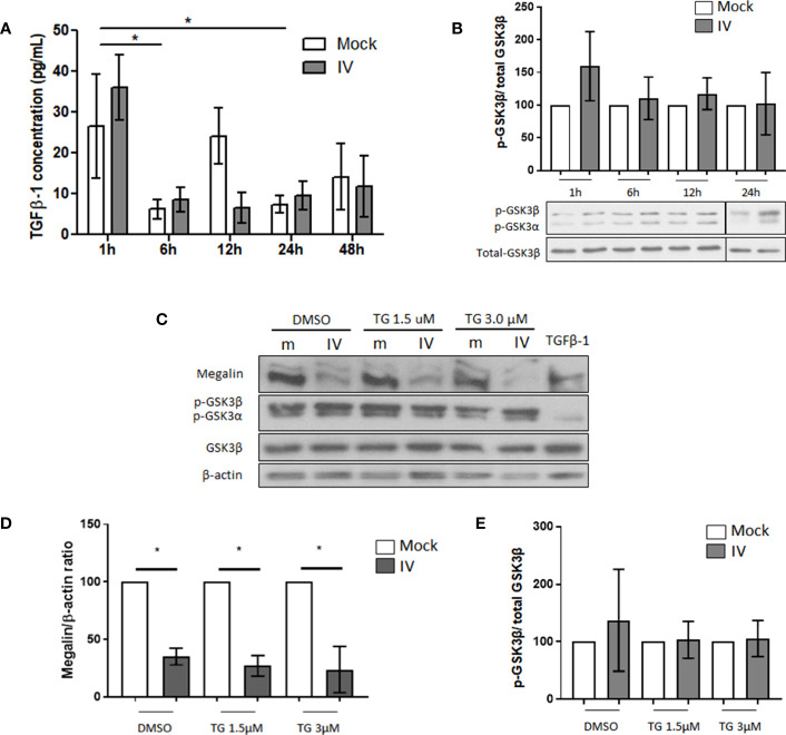
Downregulation of megalin in IV infected PCLS is independent of GSK3β activation. (A) Quantification of active TGF-β1 in media from mock and IV-inoculated PCLS incubated for 1 h, 6 h, 12 h, 24 h and 48 h (B) Phosphorylation levels of GSK3β in PCLS homogenates, at 1 h, 6 h, 12 h and 24 h post IV inoculation. (C–E) PCLS were treated with the irreversible GSK3β inhibitor tideglusib (TG) at two different concentrations (1.5 µM and 3 µM) or with a vehicle control (DMSO) for 24 h prior to inoculation with IV. Proteins were extracted and analyzed by western blot. (C) Representative immunoblots for megalin and phospho-GSK3β. TGF-β1 20 ng/mL was used as a TGF-β1 activity positive control. (D) Densitometric quantification of megalin in C. (E) Densitometric quantification of phospho-GSK3β in (C) All graphs show mean ± SD of 3 to 6 independent experiments. *p<0.05.

