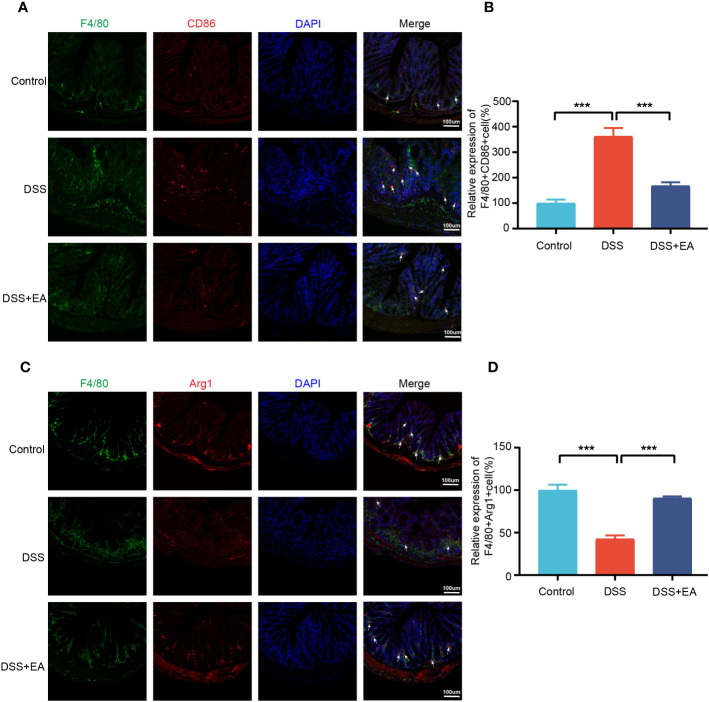
Figure 8.
Colonic macrophage polarization in the different groups. (A) Colon macrophages M1 polarizations were stained with anti-F4/80 and anti-CD86 antibodies and observed by fluorescence image (green expressed F4/80+ cells, red expressed CD86+ cells, and blue expressed DAPI; the white arrow represents F4/80+CD86+ cells). (B) The relative expression of macrophage M1 polarization (F4/80+CD86+) in three groups (n=3). (C) Colon macrophages M2 polarizations were stained with anti-F4/80 and anti-Arg1 antibodies and observed by fluorescence image (green expressed F4/80+ cells, red expressed Arg1+ cells, and blue expressed DAPI; the white arrow represents F4/80+ Arg1+ cells). (D) The relative expression of macrophage M2 polarization (F4/80+Arg1+) in three groups (n = 3). Data are show as mean ± SEM. ***P < 0.001.

