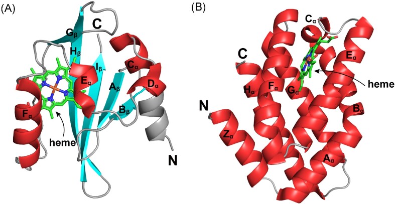
Figure 1.
Crystal structures of two types of heme-based O2 sensors. (A) Crystal structure of the heme-containing PAS domain from Escherichia coli EcDosP (PDB entry: 1V9Z) (Kurokawa et al. 2004). Its five β-strands (Aβ, Bβ, Gβ, Hβ, and Iβ) and four flanking α-helices (Cα, Dα, Eα, and Fα) are labeled as indicated. (B) Crystal structure of the heme-containing GCS domain from Escherichia coli EcDosC (PDB entry: 4ZVB) (Tarnawski et al. 2015). Each monomer contains eight α-helices, which are named Zα, Aα, Bα, Cα, Eα, Fα, Gα, and Hα according to the classical globin nomenclature. The heme ligand in each domain is indicated by arrows.

