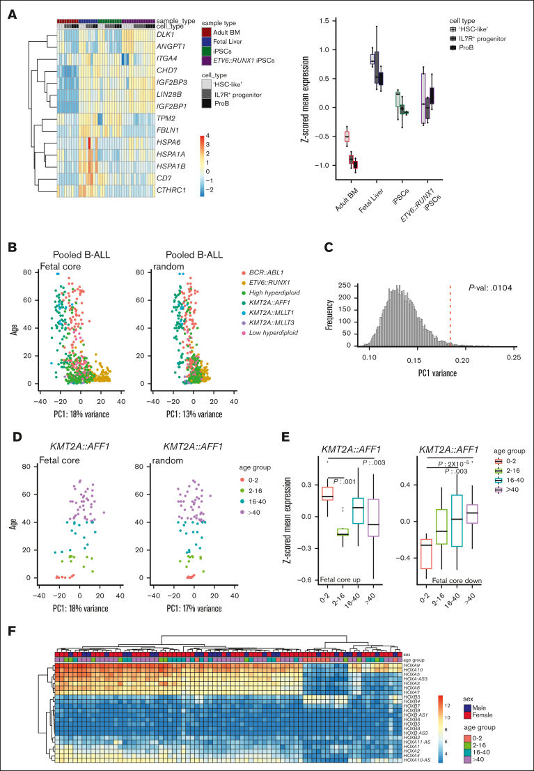
Figure 5.
The fetal core signature differentiates between pediatric and adult ALL. (A) Heatmap (left) and box plots (z-scored mean expression, right) displaying expression of fetal universally upregulated genes in an iPSC model expressing ETV6::RUNX1.15 Top rows show sample and cell types (“HSC-like,” IL7R+ progenitor, and proB). (B) PCA of pooled samples from patients with B-ALL using the fetal core signature (left) and a representative plot of random genes (right). PC1 vs age. Color coded according to translocation status. (C) Histogram showing variance of PC1 of the pooled samples with random selected genes (10 000 iterations). The fetal core PC1 variance is indicated by a dotted red line. (D) PCA of KMT2A::AFF1 (MLL::AF4) using the fetal core signature (left) and representative plot of random genes (right). PC1 vs age (color coded per age group). (E) Boxplots showing z-scored mean expression of the fetal core signature in KMT2A::AFF1 per age group. Upregulated and downregulated gene sets are shown separately, adjusted P values < 0.05 indicated in the figure. The box includes first to third quartile and the line indicates the median. (F) Heatmap of HOX gene expression in KMT2A::AFF1 B-ALL. Top rows show sex and age group, respectively.

