
FIGURE 1.
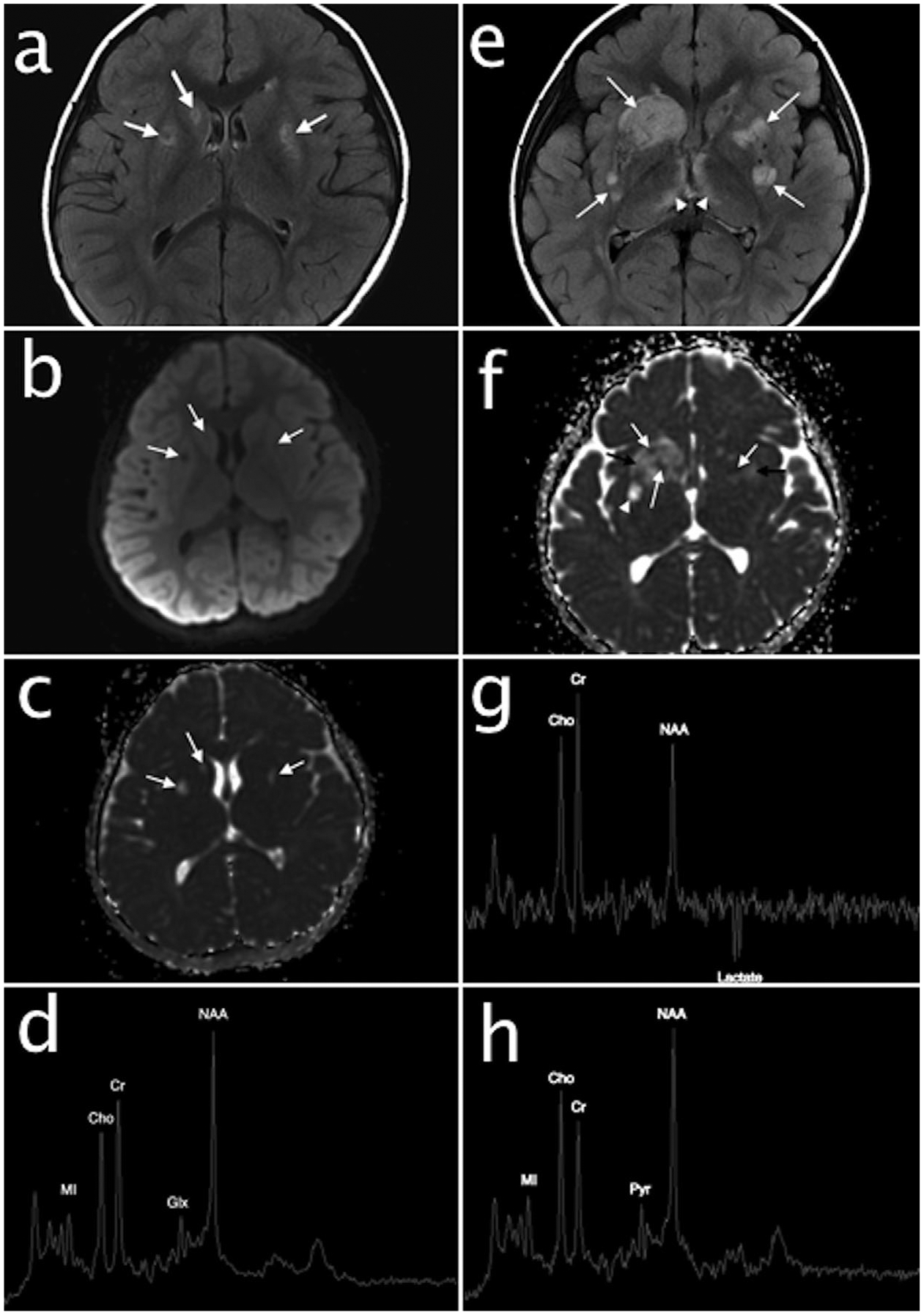
Baseline brain MRI and MRS performed at 2 years and 6 months. Axial T2 FLAIR image (a) and diffusion weighted images (b and c) demonstrates hyperintense lesions in the putamen and right caudate head with partial FLAIR signal suppression (arrows, a) and facilitated diffusion (arrows, b and c) consistent with areas of chronic encephalomalacia and necrosis. Short echo single voxel MRS (d) over the basal ganglia shows normal metabolic ratios; no abnormal pyruvate or lactate are present. Follow-up brain MRI and MRS at 3 years and 2 months. Axial T2 FLAIR (e) demonstrates acute on chronic cerebral deep gray nuclear lesions with new hyperintense lesions in the striatum (arrows) and medial thalami (arrowheads). Mixed diffusion abnormalities in the lesions (arrows, f) represent a combination of cytotoxic edema (restricted diffusion, white arrows), vasogenic edema (facilitated diffusion with mass effect, black arrows), and encephalomalacia/necrosis (facilitated diffusion without mass effect, arrowhead). Intermediate echo single voxel MRS (g) over the basal ganglia reveals an inverted lactate doublet at 1.3 ppm consistent with anaerobic metabolism and a decreased NAA to creatine ratio consistent with neuronal loss. Short echo MRS (h) over the left parietal white matter demonstrates an unusually prominent peak at 2.4 ppm consistent with pyruvate (Pyr). NAA, N-acetylaspartate; Cho, choline; Cr, creatine; MI, myoinositol