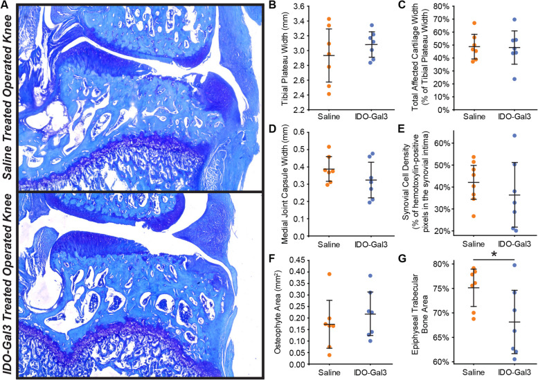
Fig. 6.
Histological scores of joint remodeling. At 12 weeks after surgery and 4 weeks after intra-articular injection, similar degrees of cartilage damage are seen in IDO-Gal3- and saline-treated knees (A). In scoring the histological images, tibial plateau width (B), total affected cartilage width (C), medial joint capsule width (D), synovial cell density (E), and osteophyte area (F) were similar for IDO-Gal3- and saline-treated groups. Epiphyseal trabecular bone area was lower in IDO-treated animals relative to saline controls, indicating marrow voids in the trabecular region were larger in IDO-Gal3-treated animals (G, p = 0.031). Bars represent mean ± 95% confidence intervals

