
Fig. 8: T cells from patients carrying PSMC3/Rpt5 variants exhibit high protein kinase R (PKR)-dependent type I IFN scores.
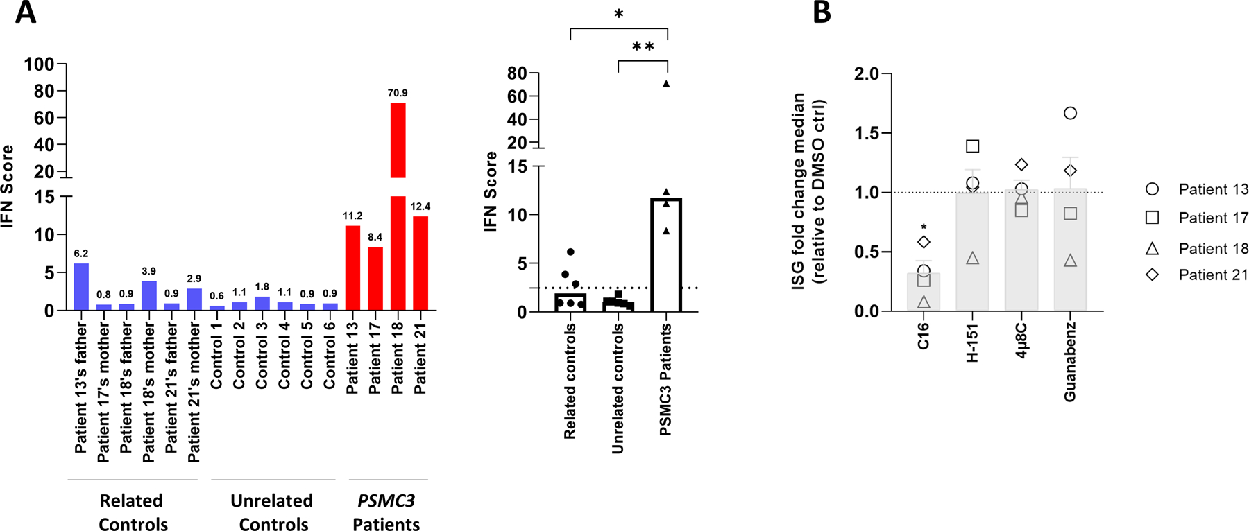
A. left panel: IFN scores for Patients #13, #17, #18 and #21 and related controls, as well as for six unrelated controls (1 to 6) were calculated as the median of the relative quantifications of the seven ISGs over a single calibrator control. Shown are the IFN scores of each sample (left panel) and the sample groups, namely parents, unrelated healthy donors and patients carrying PSMC3 variants, as indicated. right panel: Box plot of concatenated data. Statistical significance was assessed by unpaired t test where *indicates p<0.05 and ** indicates p<0.001. B. T cells isolated from individuals carrying PSMC3 variants were subjected to a 6-h treatment with DMSO (vehicle), C16 (500 nM), H-151 (2 µM), 4µ8C (100 µM) or Guanabenz (50 µM) inhibitors before RNA extraction and RT-qPCR for expression analysis of IFI27, IFI44L, IFIT1, ISG15, RSAD2, IFI44, OASL and MX1. Transcript expression was normalized to GAPDH and data are presented as the foldchange median values of the eight ISG relative to DMSO (gridline) for each patient in each treatment. Columns indicate the foldchange mean values ± SEM of the patient group (n=4) for each treatment. Statistical significance was assessed by ratio paired t test where * indicates p<0.05.