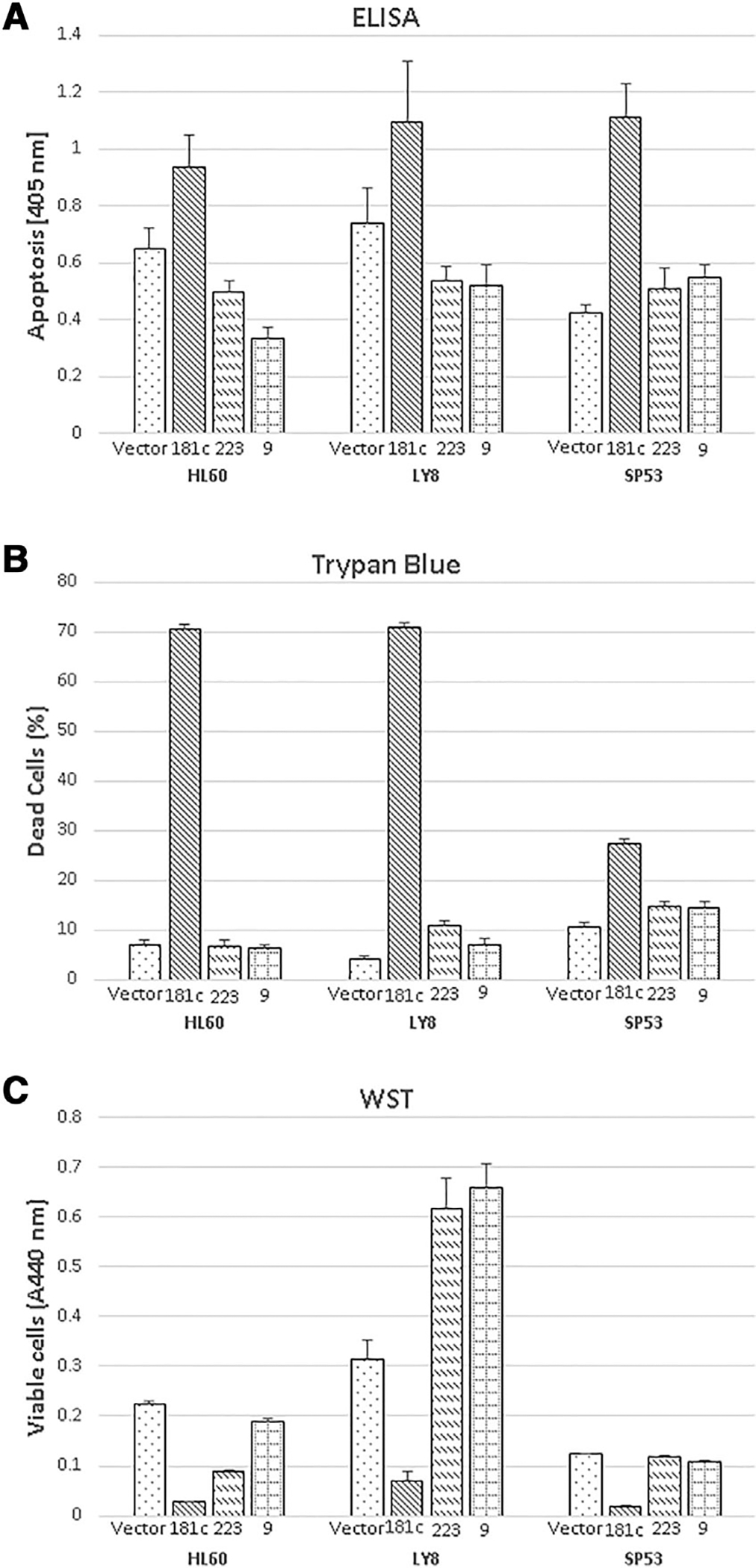
FIGURE 1. Expression of miR-181c induces apoptosis and inhibits cell proliferation.

GFP control vector, miR-181c, miR-223, and miR-9 expression plasmids were transfected into HL-60, Ly-8, and SP53 cells. Three repeated experiments were performed in triplicate. Results of technical triplicates (mean and SD) are shown. The significance is shown as compared to vector control group. *, P < 0.05; **, P < 0.01; ***, P < 0.001. A. Apoptosis was determined by Cell Death Detection ELISA 48 h after transfection. B. Dead cell percentage was obtained by trypan blue staining 60 h (HL-60 and Ly-8) or 48 h (SP53) after transfection. C. Cell proliferation was assessed by WST-1 assay 48 h after transfection
