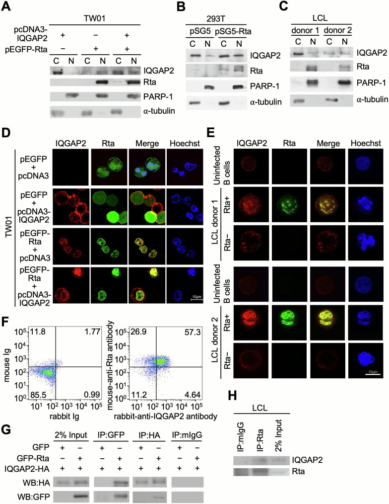
Fig 5.
IQGAP2 interacts and is colocalized with Rta in the nucleus. (A) TW01 cells were transfected with pEGFP-C1 or pEGFP-Rta, and pcDNA3 or pcDNA3-IQGAP2-HA. At 72 h post-transfection, cells were harvested for subcellular fractionation and then analyzed by western blotting for detection of IQGAP2 and Rta expression in the cytoplasm (C) and nucleus (N). (B) 293T cells were transfected with pSG5 or pSG5-Rta and subjected to subcellular fractionation at 72 h post-transfection. Expression of IQGAP2 and Rta in the cytoplasm and nucleus was analyzed by western blotting. (C) The cytoplasm and nucleus of LCLs were separated and analyzed by western blotting. Poly (ADP-ribose) polymerase-1 (PARP-1) and α-tubulin were used as nuclear and cytoplasmic markers, respectively. (D) TW01 cells were seeded onto coverslips and transfected with pcDNA3 or pcDNA3-IQGAP2-HA, and pEGFP-C1 or pEGFP-Rta. After 72 h, cells were fixed with 2% formaldehyde and permeabilized with 0.1% Triton X-100. (E) Human CD19+ uninfected B lymphocytes and EBV-immortalized LCLs were fixed with 2% formaldehyde and permeabilized with 0.1% Triton X-100. Confocal microscopy was utilized to observe the relative expression of IQGAP2 (red) and Rta (green) in the cells. Hoechst 33342 (blue) was used to indicate the nuclei of the cells. (F) LCLs were cultured in RPMI supplemented with 5% FBS for 5 d. Cells were harvested and incubated with irrelevant monoclonal mouse IgG and rabbit IgG, or mouse anti-Rta and rabbit anti-IQGAP2 antibodies. After 30 min, cells were washed with PBS and then cells were stained with fluorescein isothiocyanate (FITC)-conjugated goat anti-mouse IgG and rhodamine-conjugated goat anti-rabbit IgG antibodies for 30 min. Rta+IQGAP2+ LCLs were analyzed by flow cytometry. (G) 293T cells were transfected with both pcDNA3-IQGAP2-HA and pEGFP-C1 or pEGFP-Rta. After 72 h, cells were harvested and pre-cleared with Protein A beads. Anti-GFP antibody was used to bind GFP-Rta, while an anti-HA tag antibody was used to bind IQGAP2-HA. Protein-antibody complexes were then precipitated with Protein A beads and analyzed by western blotting to demonstrate the interaction between IQGAP2 and Rta. (H) LCLs were cultured in RPMI supplemented with 5% FBS for 5 d. Cells were harvested and pre-cleared with irrelevant monoclonal antibodies. Anti-Rta antibody was used to bind Rta. Protein-antibody complexes were then precipitated with Protein A beads and analyzed by western blotting to demonstrate the interaction between IQGAP2 and Rta.

