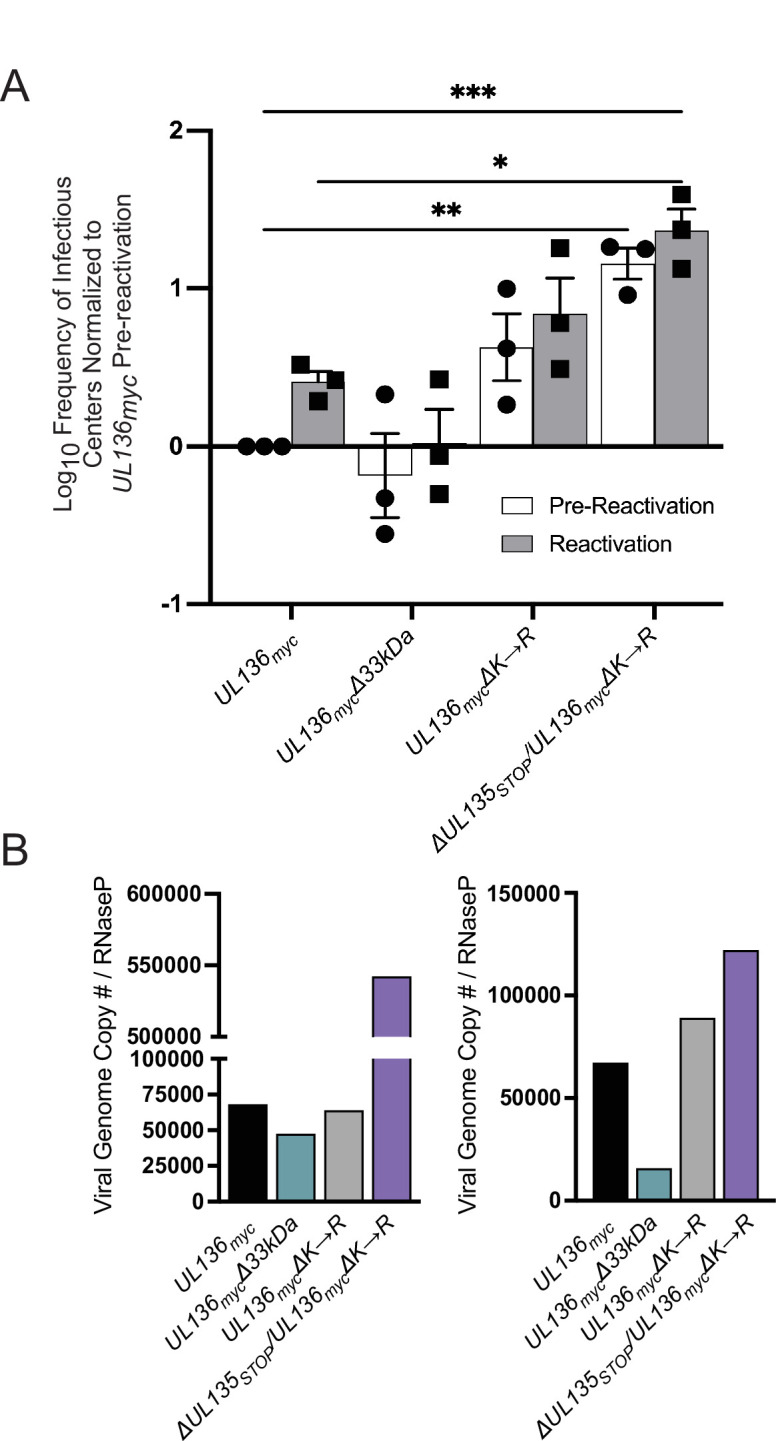
Fig 6.

Stabilizing UL136p33 rescues viral replication in the absence of UL135 in CD34+ HPCs. (A) CD34+ HPCs were infected with WT UL136myc, UL136mycΔ33kDa, UL136mycΔK→R, and ΔUL135STOP/UL136mycΔK→R at an MOI of 2. At 24 hpi, CD34+/GFP+ (infected cells) were sorted and seeded into long-term bone marrow culture. After 10 days in culture, parallel populations of either mechanically lysed cells or live cells were plated onto fibroblast monolayers in cytokine-rich media. 14 days later, GFP+ wells were scored, and the frequency of infectious centers was determined by extreme limiting dilution analysis. The mechanically lysed population defines the quantity of virus present prior to reactivation (pre-reactivation; white bar). The live-cell population defines the quantity of virus present after reactivation (reactivation; gray bar). The frequency was normalized to WT UL136myc pre-reactivation, and the average of three independent experiments is shown. Statistical significance was calculated using a two-way ANOVA with Tukey’s multiple comparison tests. *, P < 0.05; **, P < 0.01; ***, P < 0.001). (B) Total DNA was isolated from CD34+ HPCs infected with WT UL136myc, UL136mycΔ33kDa, UL136mycΔK→R, and ΔUL135STOP/UL136mycΔK→R at an MOI of 2 at 10 dpi. The number of viral genomes relative to the level of RNaseP expression was quantified by qPCR using β2.7kb RNA gene- and RNaseP-specific primers. Two biological replicates from two independent cell donors are shown.
