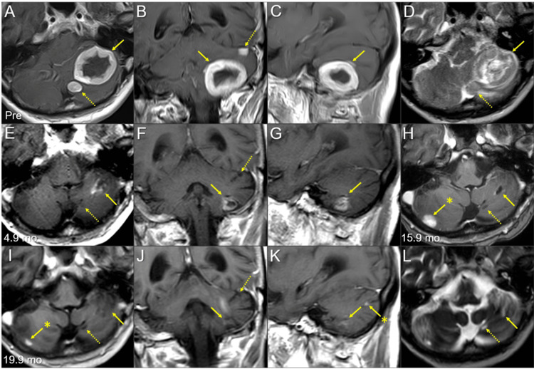
Figure 1. Magnetic resonance images of the posterior fossa lesions at the initial diagnosis and after whole-brain radiotherapy.
The images show contrast-enhanced (CE) T1-weighted images (WIs) (A-C, E-K); T2-WIs (D, L); axial images (A, D, E, H, I, L); coronal images (B, F, J); sagittal images (C, G, K); 13 days before (pre) the initiation of whole-brain radiotherapy (WBRT) (A-D); at 4.9 months (mo) after WBRT initiation (E-G); at 15.9 months (H); and at 19.9 months (I-L).
(A-L) These images are shown at the same magnification and coordinates under co-registration and fusions. (A-D) A well-demarcated lesion with the dominance of the peripheral enhancement (3.8 cm in the maximum diameter, 17.7 cm3 in the volume) (arrows in A-C) on CE-T1-WIs is observed as the heterogeneous intensity mass (arrow in D) associated with mild perilesional edema on T2-WI. The lesion reaches the cerebellar surface at the ventrolateral and caudal sides and appears to extend beyond it in some areas. In the ipsilateral hemisphere, a 1.1 cm solid lesion (0.8 cm3) is located adjacent to the 3.8 cm lesion (dashed arrow in A, D), and a 0.9 cm lesion (0.3 cm3) is located less than 1 cm away from the 3.8 cm lesion (dashed arrow in B). (E-G) At 4.9 months, T2-WIs were unavailable. The large cerebellar lesion remarkably decreased in size, along with significant attenuation of the enhancing effect (arrows in E-G), and the two adjacent lesions (dashed arrows in E, F) regressed completely. (H) At 15.9 months, the large lesion was faintly enhanced at the periphery (arrow in H). The adjacent lesion is observed as the cavitary scar without enhancement (dashed arrow in H). A new enhancing lesion (arrow with an asterisk in H) appeared. (I-L) At 19.9 months, the enhancement of the large lesion was further attenuated (arrows in I-K), with the faint high-intensity scar (arrow in L) visible on T2-WI. None of the other two lesions showed local progression (dashed arrows in I, J, and L). The right cerebellar lesion regressed markedly (arrow with an asterisk in I). A new enhancing lesion (arrow with an asterisk in K) appeared.

