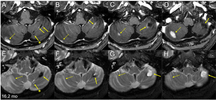
Figure 6. Multiple magnetic resonance images of the posterior fossa for the target definition of salvage stereotactic radiosurgery 16.2 months after whole-brain radiotherapy.
The images show axial CE-T1-WIs (A-D) and axial T2-WIs (E-H).
(A-H) These images are shown at the same magnification and coordinates under co-registration and fusions. Alphabetically from cranial to caudal (A-D, E-H). The large left cerebellar lesion regressed remarkably, leaving the cavitary remnant (arrows in A-H), in which the periphery was partially enhanced (arrows in A-C) and partially hypointense on T2-WIs (arrows in E, F). The adjacent medial lesion regressed completely, only leaving the tiny cavitary scar (arrows with asterisks in A and E). Two discontinuous solid-enhancing lesions (dashed arrows in A-D) developed in the right cerebellar hemisphere, which is associated with perilesional edema (dashed arrows in E-H).
mo: months; CE: contrast-enhanced; WIs: weighted images

