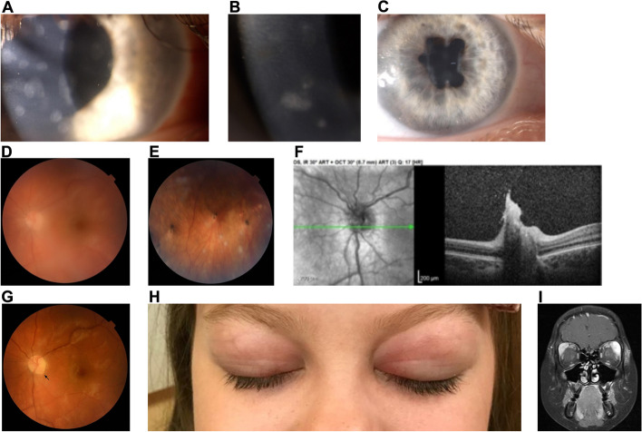
Fig. 3.
A pediatric female patient with suspected Blau Syndrome with de novo NOD2 mutation was followed for 10 years (2–12 y.o.). During the course of the disease, bilateral corneal old and new active subepithelial nummular infiltrates were noted and visualized here on color photographs of the right eye (A) and (B). The patient also developed bilateral posterior synechiae visualized here on color photograph of the right eye (C). Her fundus examination revealed bilateral vitritis and multifocal choroiditis with active and inactive peripheral lesions visualized on the left eye fundus color photography (D, E). Left eye optic disc granuloma, which appeared during relapse on adalimumab, was documented on the OCT (F) and later on the fundus color photography (G) after vitritis resolution following treatment with infliximab. Several years later, during another relapse initiated by switching to anakinra treatment, the patient developed Mikulicz’s syndrome with enlargement of bilateral lacrimal glands visualized on (H) color photography and on (I) brain MRI, coronal plane. The disease is now controlled with JAK-1 inhibitor baricitinib. Black arrow: optic disc granuloma

