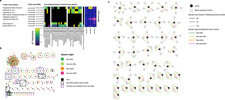
Fig. 7. Phage-host interactions and viral micro-diversity.
Based on k-mer frequency patterns, vOTUs were predicted to match host MAGs and isolate genomes (middle). Further host evidence (left) was derived from vConTACT2 viral clustering (VC) with known phages from reference database and CRISPR spacer matches from MAGs. The heatmap (right) depicts the coverage of vOTUs in the three size fractions. D2* is a dissimilarity measure (the lower, the higher the similarity) (a) CRISPR-spacer to vOTU protospacer matches at 100% similarity reveal ten clusters with slick SML derived spacers, with C1-C5 including a slick SML specific vOTU from (a), framed in purple (b). Viral micro-diversity for different water sample types and filtered size fractions. Open circles represent strain variants of the viral OTUs (closed circles) and lines indicate the sample in which the respective variant has been detected. This figure corresponds to the results shown in Table S12b. FL free-living fraction (5–0.2 µm), PA particle-associated fraction (>5 µm), SML sea-surface microlayer, SSW subsurface water, Vir viral fraction (<0.2 µm) (c).

