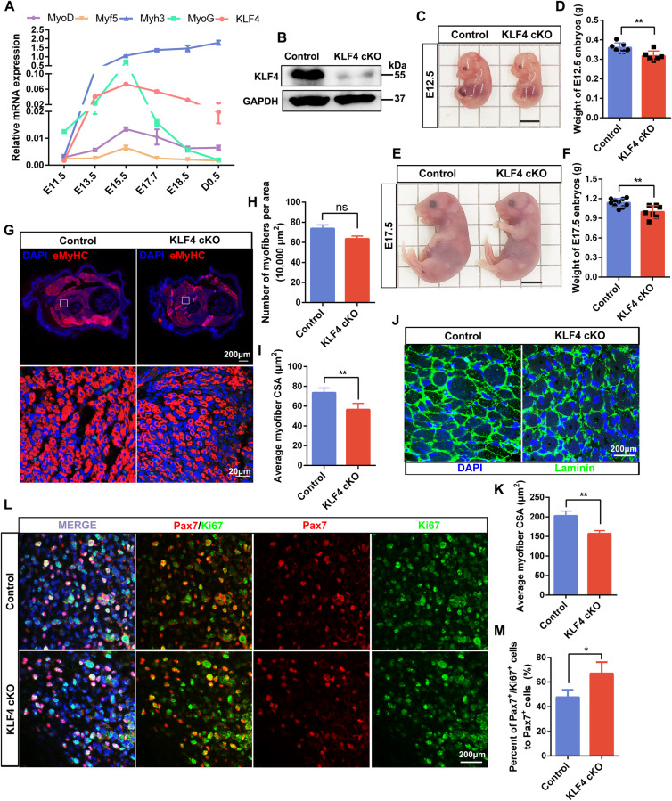
Fig. 2. KLF4 is critical for embryonic myogenesis.
A qPCR measurement of mRNA expression of KLF4 and myogenic markers (Myf5, MyoD, Myogenin, and Myh3) in the dorsal muscle of mouse at several developmental times. GAPDH was used as an internal control for normalization. E: days of embryo age, P: days of age post-birth. B WB detected the protein level of KLF4 in limbs of E12.5 embryos. C Representative images of control and KLF4 cKO embryos at E12.5. Scale bar = 0.5 cm. D Quantifications of the control and KLF4 cKO embryos weight at E12.5 (n = 7). E Representative images of control and KLF4 cKO embryos at E17.5. Scale bar = 0.5 cm. F Quantifications of the control and KLF4 cKO embryos weight at E17.5 (control: n = 10; KLF4 cKO: n = 7). G. Immunofluorescence staining of embryonic myosin heavy chain (eMyHC) was performed on cross sections of limbs of control and KLF4 cKO embryos at E12.5. Nuclei are counterstained with DAPI. Scale bar = 200 μm (top) or 20 μm (bottom). H, I. Quantifications of the numbers of eMyHC+ fibers per area and average myofiber CSA in (G)(n = 3). J Immunofluorescence staining of Laminin was performed on cross sections of limbs of control and KLF4 cKO embryos at E17.5. Scale bar = 200 μm. K Quantifications of average myofiber CSA in (J) (n = 3). L Immunofluorescence staining of Pax7 and Ki67 on cross sections of limbs of control and KLF4 cKO embryos at E12.5. Scale bar = 200 μm. M The percentage of Pax7/Ki67 double-positive cells compared with Pax7-positive cells in (L) was presented (n = 3). Data are represented as mean ± SD. *P < 0.05, **P < 0.01, ***P < 0.001; ns not significant (Student’s t test).

