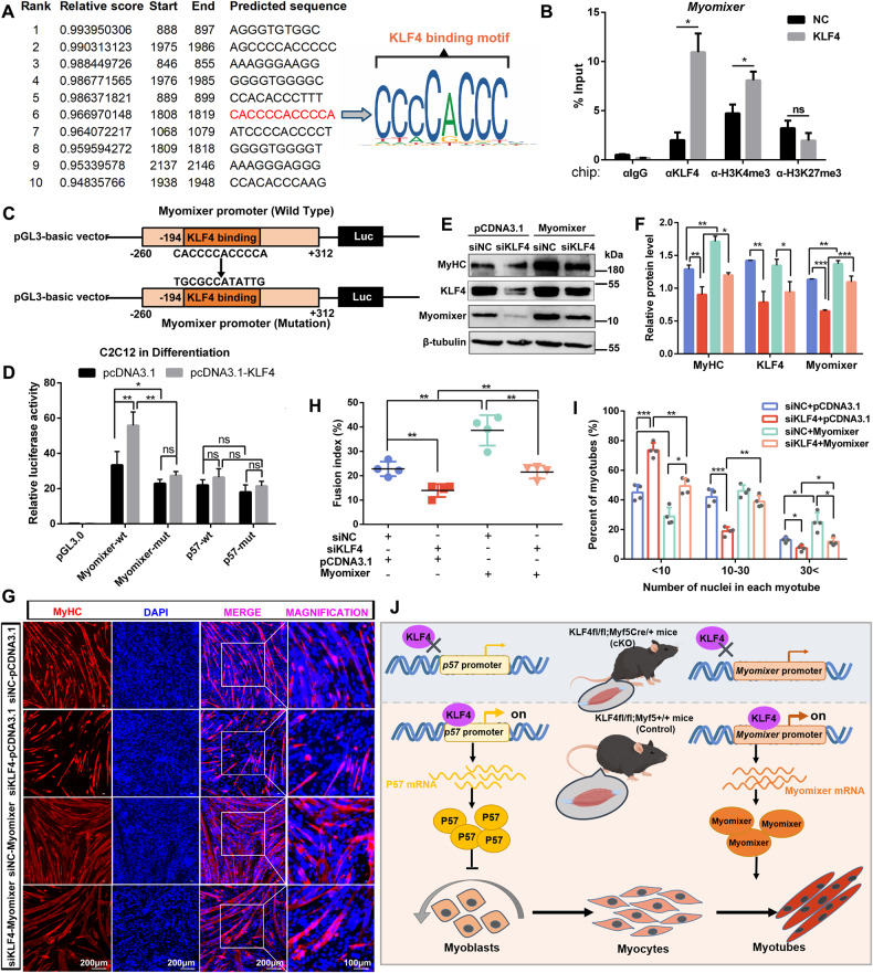
Fig. 8. KLF4 directly binds to the Myomixer promoter and activates its transcription.
A JASPAR database was used to predict KLF4 binding sites on Myomixer promoter (−2000bp ~ +300 bp, relative to the TSS of Myomixer gene). The verified sequence containing KLF4-binding motif in this study was emphasized with red. The sequence logo created in JASPAR was shown. B ChIP-qPCR analyses of KLF4, H3K4me3, and H3K27me3 enrichment on Myomixer promoter in control and KLF4 overexpression cells. Data were normalized as a percentage of the input. C Experimental design to amplify the promoter sequence of Myomixer from −260 bp to +312 bp (relative to the TSS of Myomixer gene) with the predicted KLF4 binding site was mutated, then inserted into the promoter-driven luciferase (Luc) reporter plasmid pGL3 for the dual luciferase assay. D The dual-luciferase reporter assays were performed in C2C12 cells transfected with pCDNA3.1 vector or pCDNA3.1-KLF4 vector and had differentiated for 2 d in DM, using reporter plasmids containing the wild type or mutated Myomixer promoter (n = 3). E C2C12 cells, cotransfected with si-NC or si-KLF4 and pcDNA3.1 or pcDNA3.1-Myomixer vector, were induced to differentiation in DM for 3 d. The protein levels of KLF4, MyHC, and Myomixer were detected by WB. F The relative protein levels of target proteins normalized to β-tubulin signals in (E) were obtained through WB band grey scanning. G C2C12 cells were treated as indicated in (E), and MyHC immunofluorescence staining was performed to compare myoblast fusion between four experiment groups. H, I. The fusion indexes and number of nuclei in each myotube and in (G) were quantified in six microscopic fields for each group (n = 3). J Schematic of KLF4 regulates myoblast proliferation and fusion via targeting the promoters of P57 and Myomixer. Data are represented as mean ± SD. *P < 0.05; **P < 0.01; ***P < 0.001 (Student’s t test).

