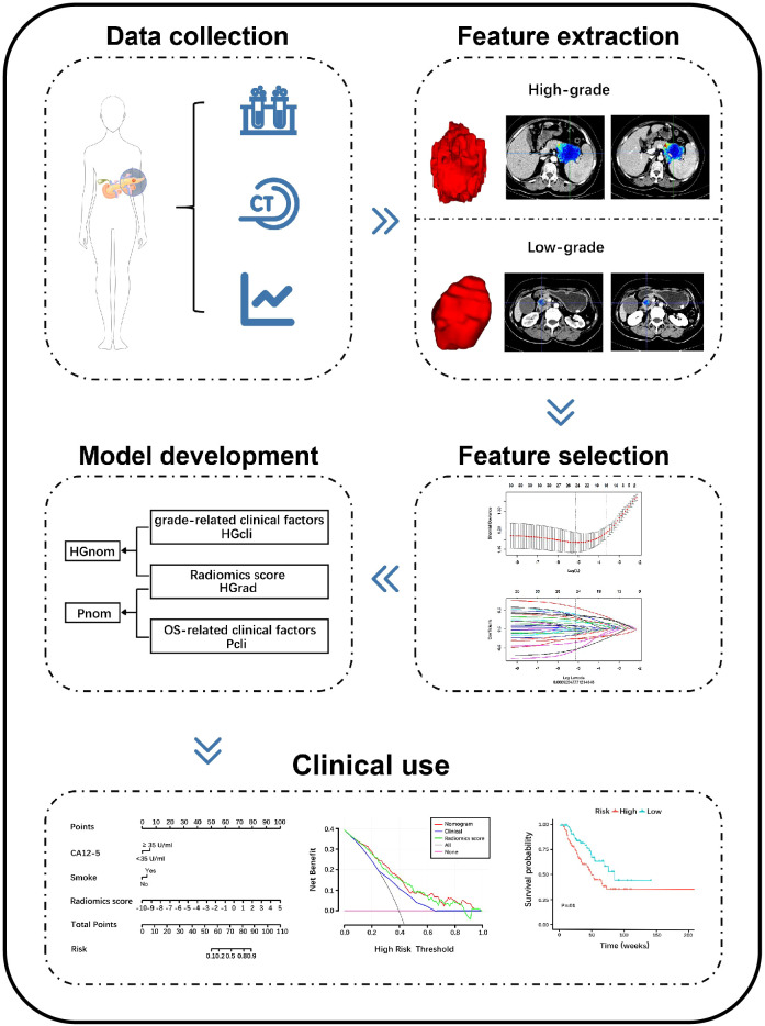
Figure 2.
Schema shows radiomics workflow. Patients data were collected. Region of interests (ROIs) were manually delineated along the entire tumor outline on all contiguous slices, and features were extracted from three-dimensional ROIs. The least absolute shrinkage and selection operator were applied to select features. The models were constructed to discriminate high-grade and low-grade PDAC and predict overall survival. The performance of the models was evaluated. PDAC, pancreatic ductal adenocarcinoma; CECT, contrast-enhanced CT; HGrad, histological grading radiomics score; HGcli, histological grading clinical model; HGnom, histological grading nomogram; Pcli, prognostic clinical model; Pnom, prognostic nomogram; CA12-5, carbohydrate antigen 12-5.

