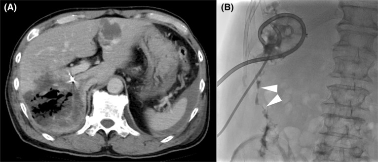
FIGURE 2.

Images following the hemostatic procedure. (A) Computed tomography on the fifth hospital day. There was gas concentrated in the necrotic area of the liver, and infection originating from the bile duct was suspected. Note the presence of a hemostatic coil in the main trunk of the posterior arterial section. (B) Necrotic cavity imaging after placement of two drainage tubes in the area of necrosis. Fistula formation (arrowheads) from the necrotic area to the colon was proven.
