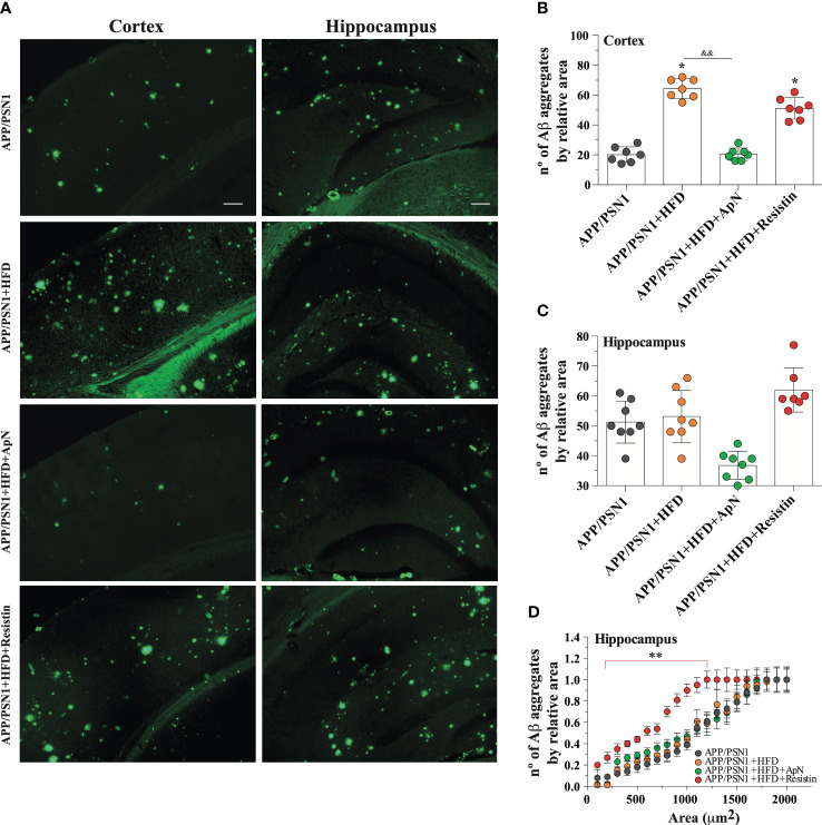
Figure 5.
ApN reduces Aβ deposition in HFD-fed APP/PSN1 mice. (A) Representative images of thioflavin S staining in the cortex or hippocampus of APP/PSN1 mice fed with either control diet or HFD in the presence of ApN or resistin. Scale bar: 200 μm. (B, C) Quantification of the number of amyloid plaques in the aforementioned conditions. (D) Quantification of the number of amyloid plaques by sectional area relative to plaque size (μm) in cortical or hippocampal slices in the aforementioned conditions. Means ± SEM are shown (n = 7-8 mice per condition in 3 technical replicates). Statistical significance was determined by one-way ANOVA, followed by Bonferroni’s multiple comparison post-hoc test. *p < 0.05 and **p < 0.01, compared with control group; &&p < 0.01, compared with specified group.

